Zinc »
PDB 4ad9-4arf »
4ajm »
Zinc in PDB 4ajm: Development of A Plate-Based Optical Biosensor Methodology to Identify PDE10 Fragment Inhibitors
Enzymatic activity of Development of A Plate-Based Optical Biosensor Methodology to Identify PDE10 Fragment Inhibitors
All present enzymatic activity of Development of A Plate-Based Optical Biosensor Methodology to Identify PDE10 Fragment Inhibitors:
3.1.4.17; 3.1.4.35;
3.1.4.17; 3.1.4.35;
Protein crystallography data
The structure of Development of A Plate-Based Optical Biosensor Methodology to Identify PDE10 Fragment Inhibitors, PDB code: 4ajm
was solved by
S.Geschwindner,
P.Johansson,
L.Spadola,
T.Akerud,
E.Back,
P.Hillertz,
R.Horsefeld,
C.Scott,
N.Spear,
G.Tian,
A.Tigerstrom,
D.Aharony,
J.S.Albert,
with X-Ray Crystallography technique. A brief refinement statistics is given in the table below:
| Resolution Low / High (Å) | 14.63 / 2.40 |
| Space group | P 21 21 21 |
| Cell size a, b, c (Å), α, β, γ (°) | 49.986, 81.694, 164.764, 90.00, 90.00, 90.00 |
| R / Rfree (%) | 21.03 / 24.51 |
Other elements in 4ajm:
The structure of Development of A Plate-Based Optical Biosensor Methodology to Identify PDE10 Fragment Inhibitors also contains other interesting chemical elements:
| Fluorine | (F) | 1 atom |
| Magnesium | (Mg) | 2 atoms |
Zinc Binding Sites:
The binding sites of Zinc atom in the Development of A Plate-Based Optical Biosensor Methodology to Identify PDE10 Fragment Inhibitors
(pdb code 4ajm). This binding sites where shown within
5.0 Angstroms radius around Zinc atom.
In total 2 binding sites of Zinc where determined in the Development of A Plate-Based Optical Biosensor Methodology to Identify PDE10 Fragment Inhibitors, PDB code: 4ajm:
Jump to Zinc binding site number: 1; 2;
In total 2 binding sites of Zinc where determined in the Development of A Plate-Based Optical Biosensor Methodology to Identify PDE10 Fragment Inhibitors, PDB code: 4ajm:
Jump to Zinc binding site number: 1; 2;
Zinc binding site 1 out of 2 in 4ajm
Go back to
Zinc binding site 1 out
of 2 in the Development of A Plate-Based Optical Biosensor Methodology to Identify PDE10 Fragment Inhibitors
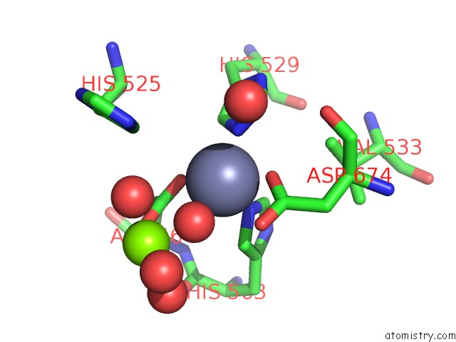
Mono view
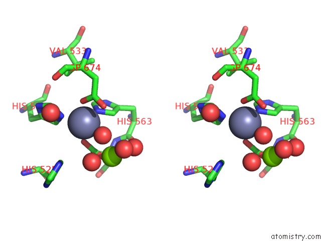
Stereo pair view
Mono view
Stereo pair view
A full contact list of Zinc with other atoms in the Zn binding
site number 1 of Development of A Plate-Based Optical Biosensor Methodology to Identify PDE10 Fragment Inhibitors within 5.0Å range:
|
Zinc binding site 2 out of 2 in 4ajm
Go back to
Zinc binding site 2 out
of 2 in the Development of A Plate-Based Optical Biosensor Methodology to Identify PDE10 Fragment Inhibitors
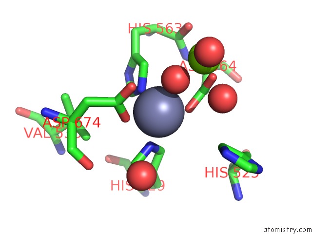
Mono view
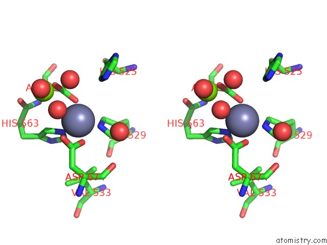
Stereo pair view
Mono view
Stereo pair view
A full contact list of Zinc with other atoms in the Zn binding
site number 2 of Development of A Plate-Based Optical Biosensor Methodology to Identify PDE10 Fragment Inhibitors within 5.0Å range:
|
Reference:
S.Geschwindner,
P.Johansson,
L.Spadola,
T.Akerud,
E.Back,
P.Hillertz,
R.Horsefeld,
C.Scott,
N.Spear,
G.Tian,
A.Tigerstrom,
D.Aharony,
J.S.Albert.
Development of A Plate-Based Optical Biosensor Methodology to Identify PDE10 Fragment Inhibitors To Be Published.
Page generated: Sat Oct 26 19:16:44 2024
Last articles
Fe in 2YXOFe in 2YRS
Fe in 2YXC
Fe in 2YNM
Fe in 2YVJ
Fe in 2YP1
Fe in 2YU2
Fe in 2YU1
Fe in 2YQB
Fe in 2YOO