Zinc »
PDB 3woj-3x0d »
3wok »
Zinc in PDB 3wok: Crystal Structure of the Dap Bii (Space)
Protein crystallography data
The structure of Crystal Structure of the Dap Bii (Space), PDB code: 3wok
was solved by
Y.Sakamoto,
Y.Suzuki,
I.Iizuka,
C.Tateoka,
S.Roppongi,
M.Fujimoto,
T.Nonaka,
W.Ogasawara,
N.Tanaka,
with X-Ray Crystallography technique. A brief refinement statistics is given in the table below:
| Resolution Low / High (Å) | 29.56 / 1.95 |
| Space group | P 21 21 21 |
| Cell size a, b, c (Å), α, β, γ (°) | 76.740, 130.440, 171.460, 90.00, 90.00, 90.00 |
| R / Rfree (%) | 18.2 / 22.5 |
Zinc Binding Sites:
The binding sites of Zinc atom in the Crystal Structure of the Dap Bii (Space)
(pdb code 3wok). This binding sites where shown within
5.0 Angstroms radius around Zinc atom.
In total 4 binding sites of Zinc where determined in the Crystal Structure of the Dap Bii (Space), PDB code: 3wok:
Jump to Zinc binding site number: 1; 2; 3; 4;
In total 4 binding sites of Zinc where determined in the Crystal Structure of the Dap Bii (Space), PDB code: 3wok:
Jump to Zinc binding site number: 1; 2; 3; 4;
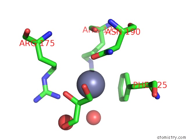
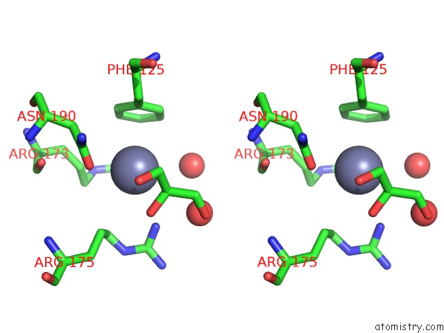
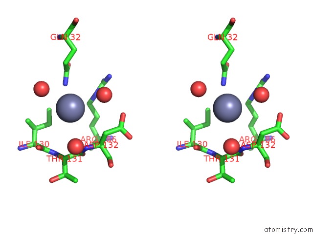
Zinc binding site 1 out of 4 in 3wok
Go back to
Zinc binding site 1 out
of 4 in the Crystal Structure of the Dap Bii (Space)
Mono view
Stereo pair view
Mono view
Stereo pair view
A full contact list of Zinc with other atoms in the Zn binding
site number 1 of Crystal Structure of the Dap Bii (Space) within 5.0Å range:
|
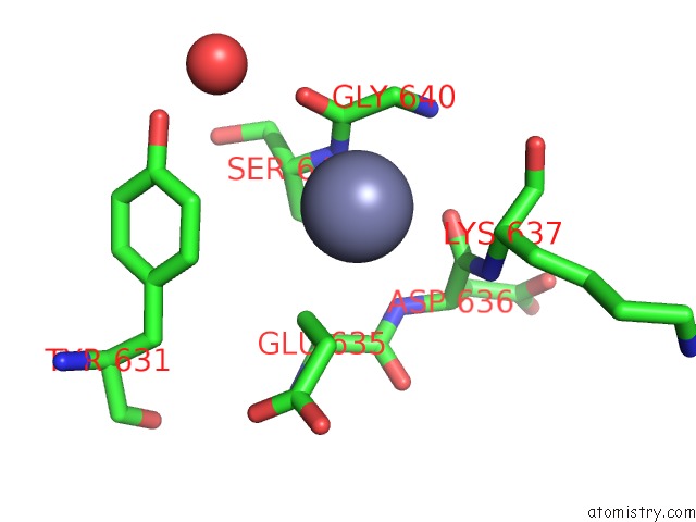
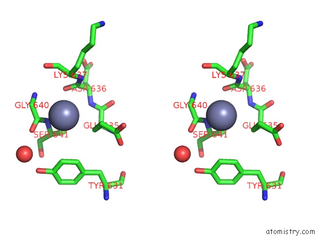
Zinc binding site 2 out of 4 in 3wok
Go back to
Zinc binding site 2 out
of 4 in the Crystal Structure of the Dap Bii (Space)
Mono view
Stereo pair view
Mono view
Stereo pair view
A full contact list of Zinc with other atoms in the Zn binding
site number 2 of Crystal Structure of the Dap Bii (Space) within 5.0Å range:
|
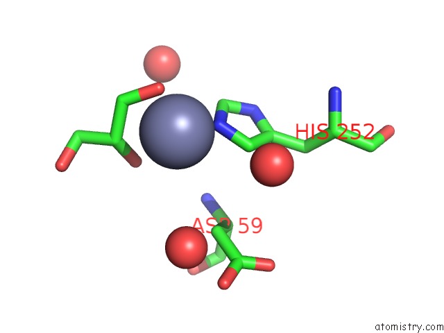
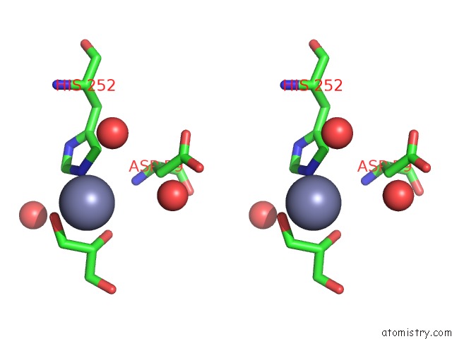
Zinc binding site 3 out of 4 in 3wok
Go back to
Zinc binding site 3 out
of 4 in the Crystal Structure of the Dap Bii (Space)

Mono view
Stereo pair view
Mono view
Stereo pair view
A full contact list of Zinc with other atoms in the Zn binding
site number 3 of Crystal Structure of the Dap Bii (Space) within 5.0Å range:
|
Zinc binding site 4 out of 4 in 3wok
Go back to
Zinc binding site 4 out
of 4 in the Crystal Structure of the Dap Bii (Space)
Mono view
Stereo pair view
Mono view
Stereo pair view
A full contact list of Zinc with other atoms in the Zn binding
site number 4 of Crystal Structure of the Dap Bii (Space) within 5.0Å range:
|
Reference:
Y.Sakamoto,
Y.Suzuki,
I.Iizuka,
C.Tateoka,
S.Roppongi,
M.Fujimoto,
K.Inaka,
H.Tanaka,
M.Masaki,
K.Ohta,
H.Okada,
T.Nonaka,
Y.Morikawa,
K.T.Nakamura,
W.Ogasawara,
N.Tanaka.
S46 Peptidases Are the First Exopeptidases to Be Members of Clan Pa Sci Rep V. 4 4977 2014.
ISSN: ESSN 2045-2322
PubMed: 24827749
DOI: 10.1038/SREP04977
Page generated: Sat Oct 26 18:12:29 2024
ISSN: ESSN 2045-2322
PubMed: 24827749
DOI: 10.1038/SREP04977
Last articles
Mn in 4M8DMn in 4MIG
Mn in 4MK5
Mn in 4MK1
Mn in 4MK2
Mn in 4M5R
Mn in 4MDA
Mn in 4MGH
Mn in 4M5U
Mn in 4M3C