Zinc »
PDB 3w5k-3woi »
3w7y »
Zinc in PDB 3w7y: 0.92A Structure of 2ZN Human Insulin at 100K
Protein crystallography data
The structure of 0.92A Structure of 2ZN Human Insulin at 100K, PDB code: 3w7y
was solved by
N.Sakabe,
K.Sakabe,
K.Sasaki,
M.Murayoshi,
with X-Ray Crystallography technique. A brief refinement statistics is given in the table below:
| Resolution Low / High (Å) | 20.00 / 0.92 |
| Space group | H 3 |
| Cell size a, b, c (Å), α, β, γ (°) | 81.120, 81.120, 33.930, 90.00, 90.00, 120.00 |
| R / Rfree (%) | 16.1 / 18 |
Zinc Binding Sites:
The binding sites of Zinc atom in the 0.92A Structure of 2ZN Human Insulin at 100K
(pdb code 3w7y). This binding sites where shown within
5.0 Angstroms radius around Zinc atom.
In total 2 binding sites of Zinc where determined in the 0.92A Structure of 2ZN Human Insulin at 100K, PDB code: 3w7y:
Jump to Zinc binding site number: 1; 2;
In total 2 binding sites of Zinc where determined in the 0.92A Structure of 2ZN Human Insulin at 100K, PDB code: 3w7y:
Jump to Zinc binding site number: 1; 2;
Zinc binding site 1 out of 2 in 3w7y
Go back to
Zinc binding site 1 out
of 2 in the 0.92A Structure of 2ZN Human Insulin at 100K
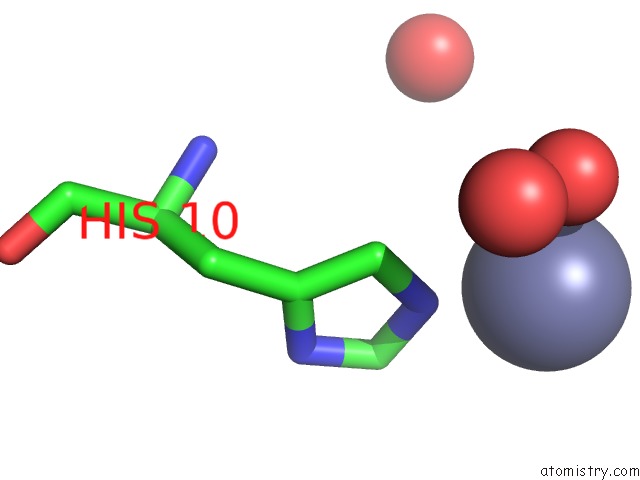
Mono view
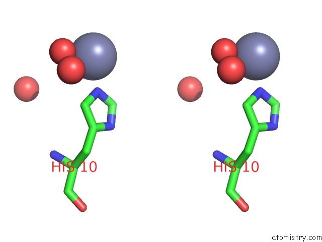
Stereo pair view
Mono view
Stereo pair view
A full contact list of Zinc with other atoms in the Zn binding
site number 1 of 0.92A Structure of 2ZN Human Insulin at 100K within 5.0Å range:
|
Zinc binding site 2 out of 2 in 3w7y
Go back to
Zinc binding site 2 out
of 2 in the 0.92A Structure of 2ZN Human Insulin at 100K
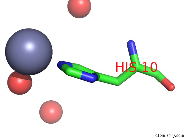
Mono view
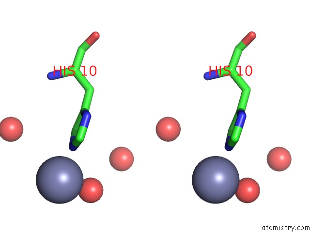
Stereo pair view
Mono view
Stereo pair view
A full contact list of Zinc with other atoms in the Zn binding
site number 2 of 0.92A Structure of 2ZN Human Insulin at 100K within 5.0Å range:
|
Reference:
N.Sakabe,
K.Sakabe,
K.Sasaki,
M.Murayoshi.
0.92A Structure of 2ZN Human Insulin at 100K To Be Published.
Page generated: Sat Oct 26 17:58:41 2024
Last articles
Fe in 7P6LFe in 7P63
Fe in 7P62
Fe in 7P61
Fe in 7P4Q
Fe in 7P46
Fe in 7P5T
Fe in 7P4M
Fe in 7P4P
Fe in 7P0P