Zinc »
PDB 3w5k-3woi »
3w6i »
Zinc in PDB 3w6i: Crystal Structure of 19F Probe-Labeled Hcai
Enzymatic activity of Crystal Structure of 19F Probe-Labeled Hcai
All present enzymatic activity of Crystal Structure of 19F Probe-Labeled Hcai:
4.2.1.1;
4.2.1.1;
Protein crystallography data
The structure of Crystal Structure of 19F Probe-Labeled Hcai, PDB code: 3w6i
was solved by
Y.Takaoka,
Y.Kioi,
A.Morito,
J.Otani,
K.Arita,
E.Ashihara,
M.Ariyoshi,
H.Tochio,
M.Shirakawa,
I.Hamachi,
with X-Ray Crystallography technique. A brief refinement statistics is given in the table below:
| Resolution Low / High (Å) | 43.35 / 2.69 |
| Space group | P 21 21 21 |
| Cell size a, b, c (Å), α, β, γ (°) | 62.463, 69.614, 119.943, 90.00, 90.00, 90.00 |
| R / Rfree (%) | 21.9 / 27 |
Other elements in 3w6i:
The structure of Crystal Structure of 19F Probe-Labeled Hcai also contains other interesting chemical elements:
| Fluorine | (F) | 12 atoms |
Zinc Binding Sites:
The binding sites of Zinc atom in the Crystal Structure of 19F Probe-Labeled Hcai
(pdb code 3w6i). This binding sites where shown within
5.0 Angstroms radius around Zinc atom.
In total 2 binding sites of Zinc where determined in the Crystal Structure of 19F Probe-Labeled Hcai, PDB code: 3w6i:
Jump to Zinc binding site number: 1; 2;
In total 2 binding sites of Zinc where determined in the Crystal Structure of 19F Probe-Labeled Hcai, PDB code: 3w6i:
Jump to Zinc binding site number: 1; 2;
Zinc binding site 1 out of 2 in 3w6i
Go back to
Zinc binding site 1 out
of 2 in the Crystal Structure of 19F Probe-Labeled Hcai

Mono view

Stereo pair view
Mono view
Stereo pair view
A full contact list of Zinc with other atoms in the Zn binding
site number 1 of Crystal Structure of 19F Probe-Labeled Hcai within 5.0Å range:
|
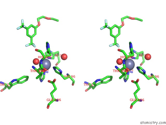
Zinc binding site 2 out of 2 in 3w6i
Go back to
Zinc binding site 2 out
of 2 in the Crystal Structure of 19F Probe-Labeled Hcai

Mono view
Stereo pair view
Mono view
Stereo pair view
A full contact list of Zinc with other atoms in the Zn binding
site number 2 of Crystal Structure of 19F Probe-Labeled Hcai within 5.0Å range:
|
Reference:
Y.Takaoka,
Y.Kioi,
A.Morito,
J.Otani,
K.Arita,
E.Ashihara,
M.Ariyoshi,
H.Tochio,
M.Shirakawa,
I.Hamachi.
Quantitative Comparison of Protein Dynamics in Live Cells and in Vitro By in-Cell 19F-uc(Nmr) To Be Published.
Page generated: Sat Oct 26 17:58:41 2024
Last articles
Fe in 7P4QFe in 7P46
Fe in 7P5T
Fe in 7P4M
Fe in 7P4P
Fe in 7P0P
Fe in 7P0R
Fe in 7P3L
Fe in 7P2C
Fe in 7P17