Zinc »
PDB 3vh4-3w5e »
3w2w »
Zinc in PDB 3w2w: Crystal Structure of the CMR2DHD-CMR3 Subcomplex Bound to Atp
Protein crystallography data
The structure of Crystal Structure of the CMR2DHD-CMR3 Subcomplex Bound to Atp, PDB code: 3w2w
was solved by
T.Numata,
T.Osawa,
with X-Ray Crystallography technique. A brief refinement statistics is given in the table below:
| Resolution Low / High (Å) | 46.91 / 2.50 |
| Space group | I 2 2 2 |
| Cell size a, b, c (Å), α, β, γ (°) | 103.579, 135.804, 191.323, 90.00, 90.00, 90.00 |
| R / Rfree (%) | 21.1 / 24.6 |
Other elements in 3w2w:
The structure of Crystal Structure of the CMR2DHD-CMR3 Subcomplex Bound to Atp also contains other interesting chemical elements:
| Magnesium | (Mg) | 2 atoms |
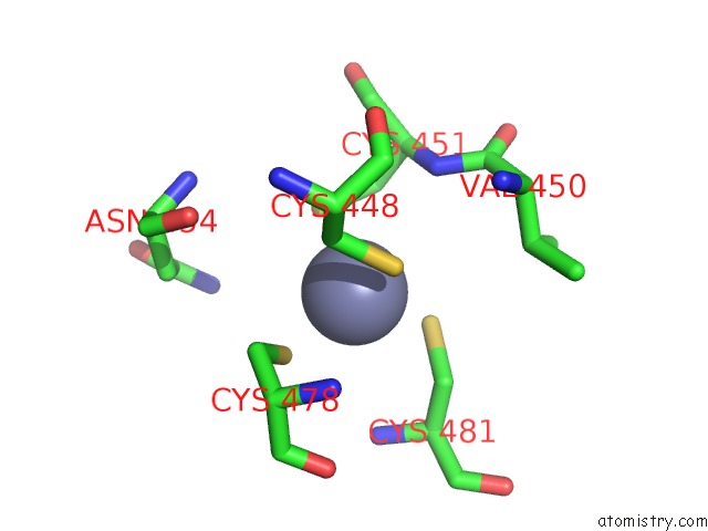
Zinc Binding Sites:
The binding sites of Zinc atom in the Crystal Structure of the CMR2DHD-CMR3 Subcomplex Bound to Atp
(pdb code 3w2w). This binding sites where shown within
5.0 Angstroms radius around Zinc atom.
In total only one binding site of Zinc was determined in the Crystal Structure of the CMR2DHD-CMR3 Subcomplex Bound to Atp, PDB code: 3w2w:
In total only one binding site of Zinc was determined in the Crystal Structure of the CMR2DHD-CMR3 Subcomplex Bound to Atp, PDB code: 3w2w:
Zinc binding site 1 out of 1 in 3w2w
Go back to
Zinc binding site 1 out
of 1 in the Crystal Structure of the CMR2DHD-CMR3 Subcomplex Bound to Atp
Mono view
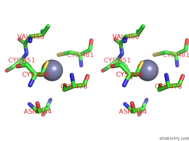
Stereo pair view
Mono view
Stereo pair view
A full contact list of Zinc with other atoms in the Zn binding
site number 1 of Crystal Structure of the CMR2DHD-CMR3 Subcomplex Bound to Atp within 5.0Å range:
|
Reference:
T.Osawa,
H.Inanaga,
T.Numata.
Crystal Structure of the CMR2-CMR3 Subcomplex in the Crispr-Cas Rna Silencing Effector Complex. J.Mol.Biol. V. 425 3811 2013.
ISSN: ISSN 0022-2836
PubMed: 23583914
DOI: 10.1016/J.JMB.2013.03.042
Page generated: Sat Oct 26 17:57:30 2024
ISSN: ISSN 0022-2836
PubMed: 23583914
DOI: 10.1016/J.JMB.2013.03.042
Last articles
Mg in 3E35Mg in 3E1F
Mg in 3E2D
Mg in 3DYP
Mg in 3DYO
Mg in 3E27
Mg in 3E25
Mg in 3E22
Mg in 3DYM
Mg in 3E18