Zinc »
PDB 3uk4-3v38 »
3uwb »
Zinc in PDB 3uwb: Crystal Structure of A Probable Peptide Deformylase From Strucynechococcus Phage S-SSM7 in Complex with Actinonin
Protein crystallography data
The structure of Crystal Structure of A Probable Peptide Deformylase From Strucynechococcus Phage S-SSM7 in Complex with Actinonin, PDB code: 3uwb
was solved by
D.Lorimer,
J.Abendroth,
T.E.Edwards,
A.Burgin,
A.Segall,
F.Rohwer,
with X-Ray Crystallography technique. A brief refinement statistics is given in the table below:
| Resolution Low / High (Å) | 42.62 / 1.70 |
| Space group | P 21 21 21 |
| Cell size a, b, c (Å), α, β, γ (°) | 47.810, 59.150, 61.460, 90.00, 90.00, 90.00 |
| R / Rfree (%) | 15.5 / 17.1 |
Other elements in 3uwb:
The structure of Crystal Structure of A Probable Peptide Deformylase From Strucynechococcus Phage S-SSM7 in Complex with Actinonin also contains other interesting chemical elements:
| Chlorine | (Cl) | 1 atom |
Zinc Binding Sites:
The binding sites of Zinc atom in the Crystal Structure of A Probable Peptide Deformylase From Strucynechococcus Phage S-SSM7 in Complex with Actinonin
(pdb code 3uwb). This binding sites where shown within
5.0 Angstroms radius around Zinc atom.
In total only one binding site of Zinc was determined in the Crystal Structure of A Probable Peptide Deformylase From Strucynechococcus Phage S-SSM7 in Complex with Actinonin, PDB code: 3uwb:
In total only one binding site of Zinc was determined in the Crystal Structure of A Probable Peptide Deformylase From Strucynechococcus Phage S-SSM7 in Complex with Actinonin, PDB code: 3uwb:
Zinc binding site 1 out of 1 in 3uwb
Go back to
Zinc binding site 1 out
of 1 in the Crystal Structure of A Probable Peptide Deformylase From Strucynechococcus Phage S-SSM7 in Complex with Actinonin
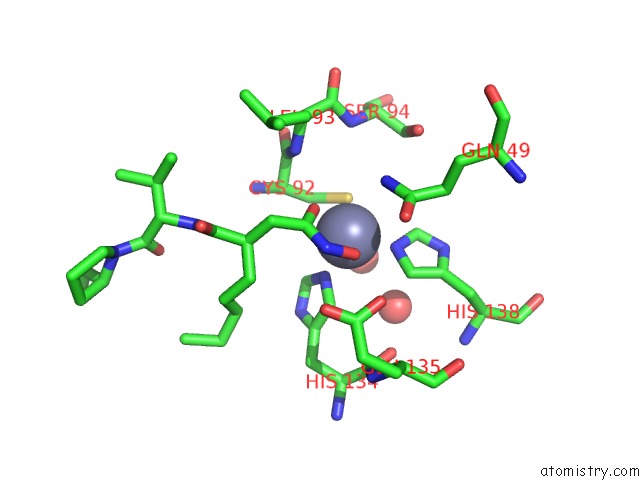
Mono view
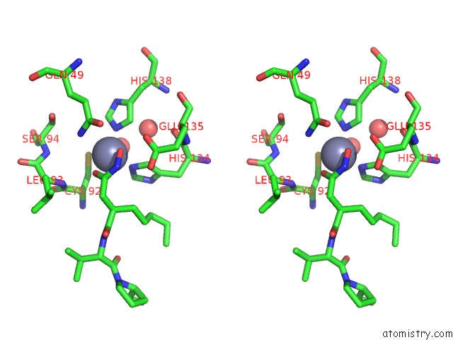
Stereo pair view
Mono view
Stereo pair view
A full contact list of Zinc with other atoms in the Zn binding
site number 1 of Crystal Structure of A Probable Peptide Deformylase From Strucynechococcus Phage S-SSM7 in Complex with Actinonin within 5.0Å range:
|
Reference:
J.A.Frank,
D.Lorimer,
M.Youle,
P.Witte,
T.Craig,
J.Abendroth,
F.Rohwer,
R.A.Edwards,
A.M.Segall,
A.B.Burgin.
Structure and Function of A Cyanophage-Encoded Peptide Deformylase. Isme J V. 7 1150 2013.
ISSN: ISSN 1751-7362
PubMed: 23407310
DOI: 10.1038/ISMEJ.2013.4
Page generated: Sat Oct 26 17:29:54 2024
ISSN: ISSN 1751-7362
PubMed: 23407310
DOI: 10.1038/ISMEJ.2013.4
Last articles
Fe in 2YXOFe in 2YRS
Fe in 2YXC
Fe in 2YNM
Fe in 2YVJ
Fe in 2YP1
Fe in 2YU2
Fe in 2YU1
Fe in 2YQB
Fe in 2YOO