Zinc »
PDB 3u6q-3uec »
3u9y »
Zinc in PDB 3u9y: Crystal Structure of Human Tankyrase 2 Catalytic Domain in Complex with Olaparib
Enzymatic activity of Crystal Structure of Human Tankyrase 2 Catalytic Domain in Complex with Olaparib
All present enzymatic activity of Crystal Structure of Human Tankyrase 2 Catalytic Domain in Complex with Olaparib:
2.4.2.30;
2.4.2.30;
Protein crystallography data
The structure of Crystal Structure of Human Tankyrase 2 Catalytic Domain in Complex with Olaparib, PDB code: 3u9y
was solved by
M.Narwal,
L.Lehtio,
with X-Ray Crystallography technique. A brief refinement statistics is given in the table below:
| Resolution Low / High (Å) | 43.61 / 2.30 |
| Space group | P 41 21 2 |
| Cell size a, b, c (Å), α, β, γ (°) | 66.560, 66.560, 115.920, 90.00, 90.00, 90.00 |
| R / Rfree (%) | 18.1 / 23.4 |
Other elements in 3u9y:
The structure of Crystal Structure of Human Tankyrase 2 Catalytic Domain in Complex with Olaparib also contains other interesting chemical elements:
| Fluorine | (F) | 1 atom |
Zinc Binding Sites:
The binding sites of Zinc atom in the Crystal Structure of Human Tankyrase 2 Catalytic Domain in Complex with Olaparib
(pdb code 3u9y). This binding sites where shown within
5.0 Angstroms radius around Zinc atom.
In total only one binding site of Zinc was determined in the Crystal Structure of Human Tankyrase 2 Catalytic Domain in Complex with Olaparib, PDB code: 3u9y:
In total only one binding site of Zinc was determined in the Crystal Structure of Human Tankyrase 2 Catalytic Domain in Complex with Olaparib, PDB code: 3u9y:
Zinc binding site 1 out of 1 in 3u9y
Go back to
Zinc binding site 1 out
of 1 in the Crystal Structure of Human Tankyrase 2 Catalytic Domain in Complex with Olaparib
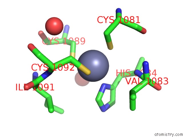
Mono view
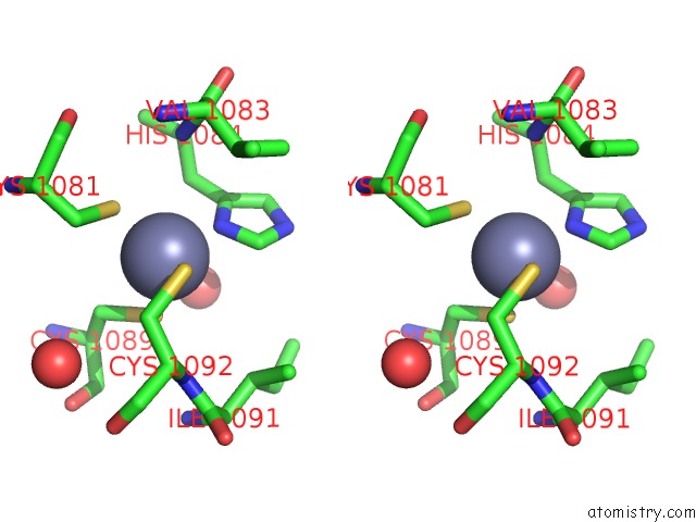
Stereo pair view
Mono view
Stereo pair view
A full contact list of Zinc with other atoms in the Zn binding
site number 1 of Crystal Structure of Human Tankyrase 2 Catalytic Domain in Complex with Olaparib within 5.0Å range:
|
Reference:
M.Narwal,
H.Venkannagari,
L.Lehtio.
Structural Basis of Selective Inhibition of Human Tankyrases. J.Med.Chem. V. 55 1360 2012.
ISSN: ISSN 0022-2623
PubMed: 22233320
DOI: 10.1021/JM201510P
Page generated: Sat Oct 26 17:04:47 2024
ISSN: ISSN 0022-2623
PubMed: 22233320
DOI: 10.1021/JM201510P
Last articles
Mg in 1L9ZMg in 1L9U
Mg in 1L8P
Mg in 1L9B
Mg in 1L8T
Mg in 1L8A
Mg in 1L8Q
Mg in 1L7N
Mg in 1L7M
Mg in 1L6Y