Zinc »
PDB 3u6q-3uec »
3u7c »
Zinc in PDB 3u7c: Crystal Structure of the V143I Mutant of Human Carbonic Anhydrase II
Enzymatic activity of Crystal Structure of the V143I Mutant of Human Carbonic Anhydrase II
All present enzymatic activity of Crystal Structure of the V143I Mutant of Human Carbonic Anhydrase II:
4.2.1.1;
4.2.1.1;
Protein crystallography data
The structure of Crystal Structure of the V143I Mutant of Human Carbonic Anhydrase II, PDB code: 3u7c
was solved by
D.M.West,
C.U.Kim,
A.H.Robbins,
R.Mckenna,
with X-Ray Crystallography technique. A brief refinement statistics is given in the table below:
| Resolution Low / High (Å) | 20.00 / 0.93 |
| Space group | P 1 21 1 |
| Cell size a, b, c (Å), α, β, γ (°) | 42.296, 41.458, 72.083, 90.00, 104.16, 90.00 |
| R / Rfree (%) | 10.5 / 13.1 |
Zinc Binding Sites:
The binding sites of Zinc atom in the Crystal Structure of the V143I Mutant of Human Carbonic Anhydrase II
(pdb code 3u7c). This binding sites where shown within
5.0 Angstroms radius around Zinc atom.
In total only one binding site of Zinc was determined in the Crystal Structure of the V143I Mutant of Human Carbonic Anhydrase II, PDB code: 3u7c:
In total only one binding site of Zinc was determined in the Crystal Structure of the V143I Mutant of Human Carbonic Anhydrase II, PDB code: 3u7c:
Zinc binding site 1 out of 1 in 3u7c
Go back to
Zinc binding site 1 out
of 1 in the Crystal Structure of the V143I Mutant of Human Carbonic Anhydrase II
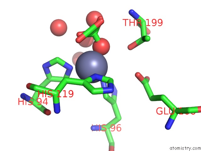
Mono view
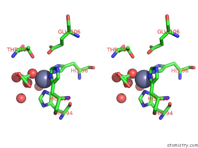
Stereo pair view
Mono view
Stereo pair view
A full contact list of Zinc with other atoms in the Zn binding
site number 1 of Crystal Structure of the V143I Mutant of Human Carbonic Anhydrase II within 5.0Å range:
|
Reference:
D.M.West,
C.U.Kim,
C.Tu,
J.Gordon,
A.H.Robbins,
S.M.Gruner,
D.M.Silverman,
R.Mckenna.
Crystal Structure of the V143I Mutant of Human Carbonic Anhydrase II To Be Published.
Page generated: Sat Oct 26 17:02:05 2024
Last articles
Mg in 1R2QMg in 1R2C
Mg in 1R10
Mg in 1R0Z
Mg in 1R0X
Mg in 1R0A
Mg in 1R03
Mg in 1QZR
Mg in 1QU2
Mg in 1QYF