Zinc »
PDB 3tus-3u6p »
3u04 »
Zinc in PDB 3u04: Crystal Structure of Peptide Deformylase From Ehrlichia Chaffeensis in Complex with Actinonin
Enzymatic activity of Crystal Structure of Peptide Deformylase From Ehrlichia Chaffeensis in Complex with Actinonin
All present enzymatic activity of Crystal Structure of Peptide Deformylase From Ehrlichia Chaffeensis in Complex with Actinonin:
3.5.1.88;
3.5.1.88;
Protein crystallography data
The structure of Crystal Structure of Peptide Deformylase From Ehrlichia Chaffeensis in Complex with Actinonin, PDB code: 3u04
was solved by
Seattle Structural Genomics Center For Infectious Disease (Ssgcid),
with X-Ray Crystallography technique. A brief refinement statistics is given in the table below:
| Resolution Low / High (Å) | 41.94 / 1.70 |
| Space group | C 1 2 1 |
| Cell size a, b, c (Å), α, β, γ (°) | 83.890, 33.020, 68.140, 90.00, 91.17, 90.00 |
| R / Rfree (%) | 17 / 19.8 |
Other elements in 3u04:
The structure of Crystal Structure of Peptide Deformylase From Ehrlichia Chaffeensis in Complex with Actinonin also contains other interesting chemical elements:
| Chlorine | (Cl) | 2 atoms |
Zinc Binding Sites:
The binding sites of Zinc atom in the Crystal Structure of Peptide Deformylase From Ehrlichia Chaffeensis in Complex with Actinonin
(pdb code 3u04). This binding sites where shown within
5.0 Angstroms radius around Zinc atom.
In total only one binding site of Zinc was determined in the Crystal Structure of Peptide Deformylase From Ehrlichia Chaffeensis in Complex with Actinonin, PDB code: 3u04:
In total only one binding site of Zinc was determined in the Crystal Structure of Peptide Deformylase From Ehrlichia Chaffeensis in Complex with Actinonin, PDB code: 3u04:
Zinc binding site 1 out of 1 in 3u04
Go back to
Zinc binding site 1 out
of 1 in the Crystal Structure of Peptide Deformylase From Ehrlichia Chaffeensis in Complex with Actinonin
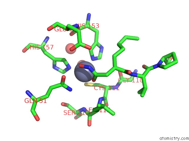
Mono view
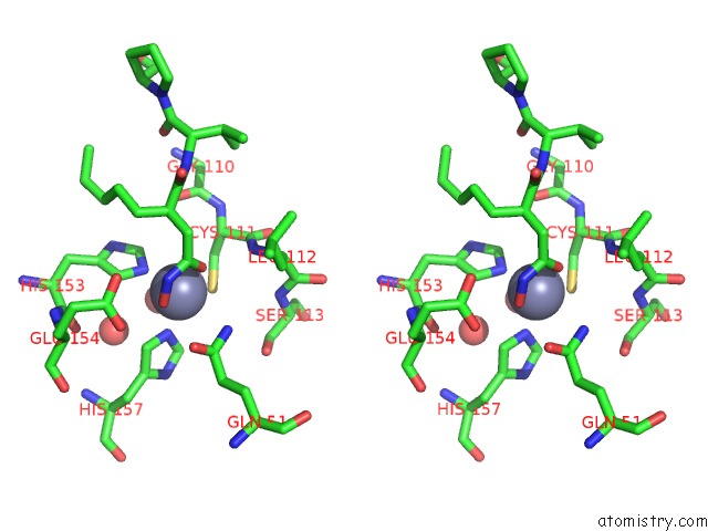
Stereo pair view
Mono view
Stereo pair view
A full contact list of Zinc with other atoms in the Zn binding
site number 1 of Crystal Structure of Peptide Deformylase From Ehrlichia Chaffeensis in Complex with Actinonin within 5.0Å range:
|
Reference:
Seattle Structural Genomics Center For Infectious Disease(Ssgcid),
J.Abendroth,
M.C.Clifton,
T.E.Edwards,
B.L.Staker.
Crystal Structure of Peptide Deformylase From Ehrlichia Chaffeensis in Complex with Actinonin To Be Published.
Page generated: Sat Oct 26 16:50:37 2024
Last articles
Mg in 1R2QMg in 1R2C
Mg in 1R10
Mg in 1R0Z
Mg in 1R0X
Mg in 1R0A
Mg in 1R03
Mg in 1QZR
Mg in 1QU2
Mg in 1QYF