Zinc »
PDB 3sck-3sjd »
3siq »
Zinc in PDB 3siq: Crystal Structure of Autoinhibited DIAP1-BIR1 Domain
Protein crystallography data
The structure of Crystal Structure of Autoinhibited DIAP1-BIR1 Domain, PDB code: 3siq
was solved by
X.Li,
J.Wang,
Y.Shi,
with X-Ray Crystallography technique. A brief refinement statistics is given in the table below:
| Resolution Low / High (Å) | 32.98 / 2.40 |
| Space group | P 31 |
| Cell size a, b, c (Å), α, β, γ (°) | 99.782, 99.782, 71.344, 90.00, 90.00, 120.00 |
| R / Rfree (%) | 14.6 / 21.7 |
Zinc Binding Sites:
The binding sites of Zinc atom in the Crystal Structure of Autoinhibited DIAP1-BIR1 Domain
(pdb code 3siq). This binding sites where shown within
5.0 Angstroms radius around Zinc atom.
In total 6 binding sites of Zinc where determined in the Crystal Structure of Autoinhibited DIAP1-BIR1 Domain, PDB code: 3siq:
Jump to Zinc binding site number: 1; 2; 3; 4; 5; 6;
In total 6 binding sites of Zinc where determined in the Crystal Structure of Autoinhibited DIAP1-BIR1 Domain, PDB code: 3siq:
Jump to Zinc binding site number: 1; 2; 3; 4; 5; 6;
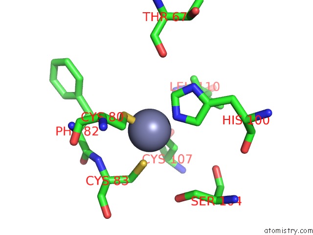
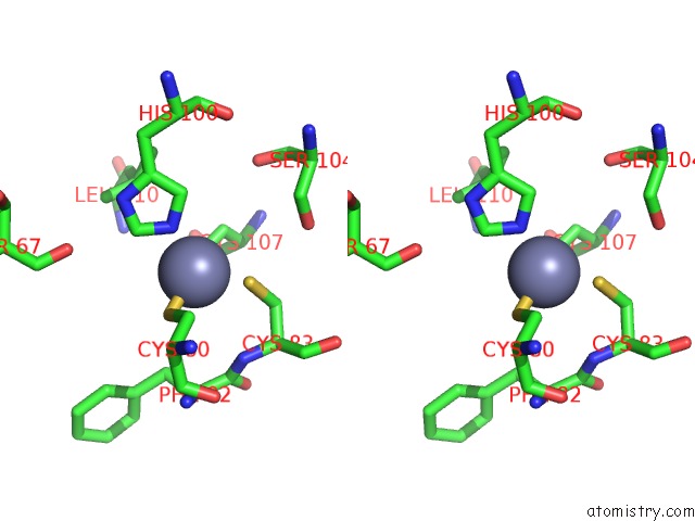
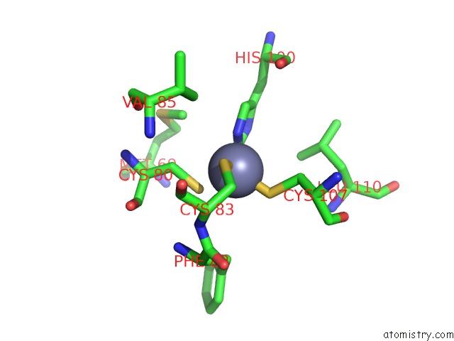
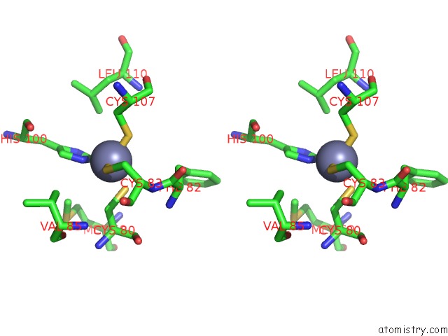
Zinc binding site 1 out of 6 in 3siq
Go back to
Zinc binding site 1 out
of 6 in the Crystal Structure of Autoinhibited DIAP1-BIR1 Domain
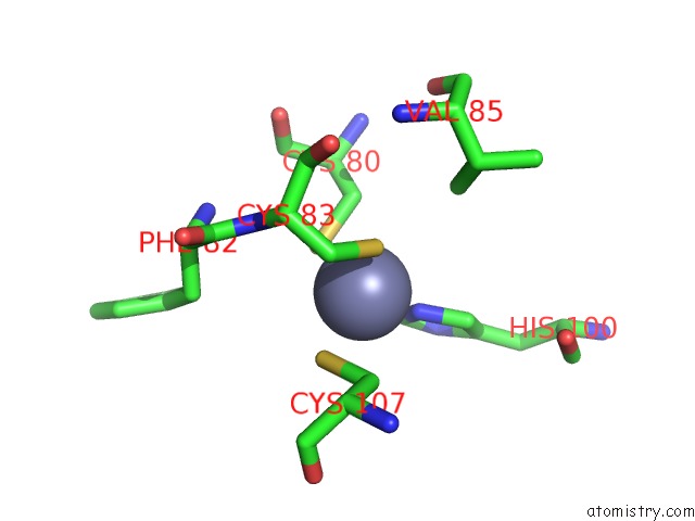
Mono view
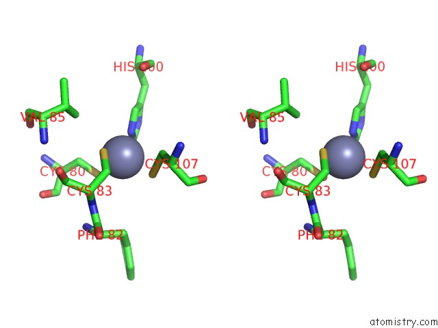
Stereo pair view
Mono view
Stereo pair view
A full contact list of Zinc with other atoms in the Zn binding
site number 1 of Crystal Structure of Autoinhibited DIAP1-BIR1 Domain within 5.0Å range:
|
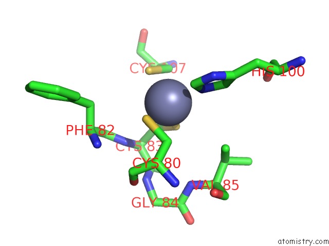
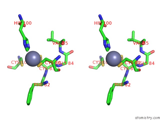
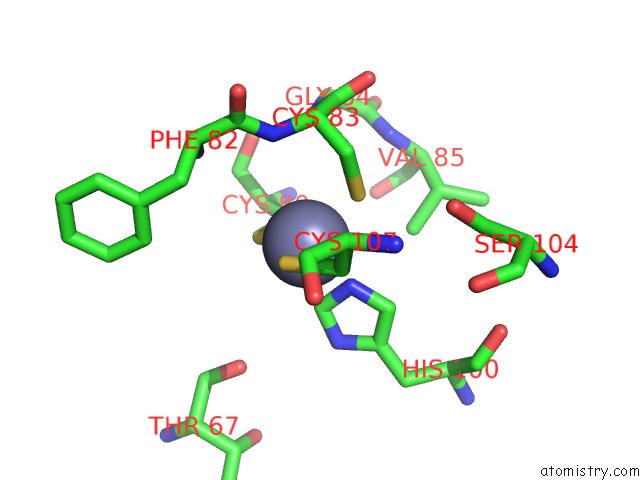
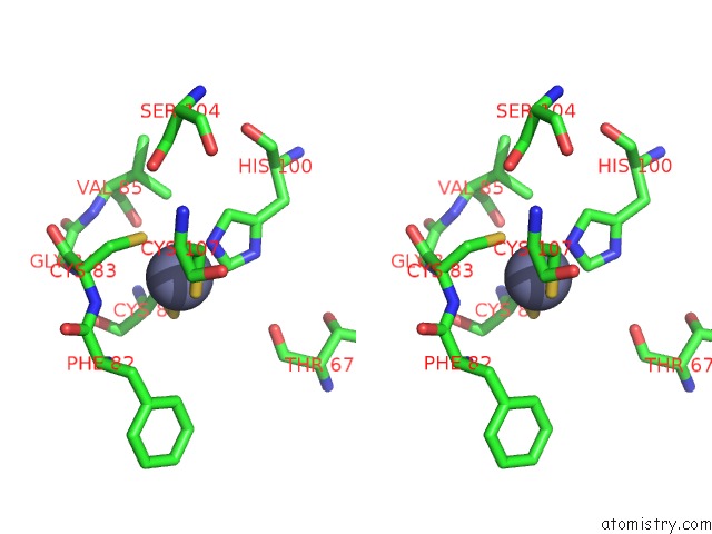
Zinc binding site 2 out of 6 in 3siq
Go back to
Zinc binding site 2 out
of 6 in the Crystal Structure of Autoinhibited DIAP1-BIR1 Domain
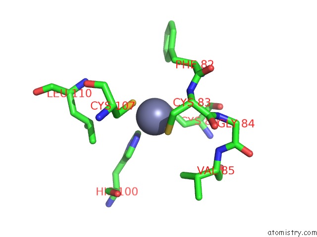
Mono view
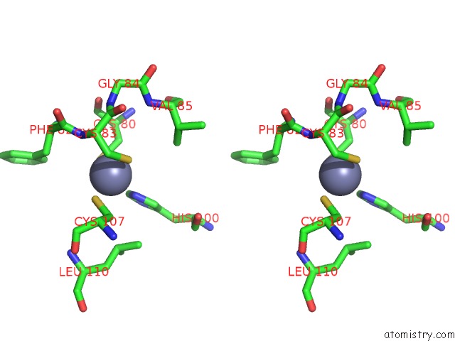
Stereo pair view
Mono view
Stereo pair view
A full contact list of Zinc with other atoms in the Zn binding
site number 2 of Crystal Structure of Autoinhibited DIAP1-BIR1 Domain within 5.0Å range:
|
Zinc binding site 3 out of 6 in 3siq
Go back to
Zinc binding site 3 out
of 6 in the Crystal Structure of Autoinhibited DIAP1-BIR1 Domain
Mono view
Stereo pair view
Mono view
Stereo pair view
A full contact list of Zinc with other atoms in the Zn binding
site number 3 of Crystal Structure of Autoinhibited DIAP1-BIR1 Domain within 5.0Å range:
|
Zinc binding site 4 out of 6 in 3siq
Go back to
Zinc binding site 4 out
of 6 in the Crystal Structure of Autoinhibited DIAP1-BIR1 Domain
Mono view
Stereo pair view
Mono view
Stereo pair view
A full contact list of Zinc with other atoms in the Zn binding
site number 4 of Crystal Structure of Autoinhibited DIAP1-BIR1 Domain within 5.0Å range:
|
Zinc binding site 5 out of 6 in 3siq
Go back to
Zinc binding site 5 out
of 6 in the Crystal Structure of Autoinhibited DIAP1-BIR1 Domain
Mono view
Stereo pair view
Mono view
Stereo pair view
A full contact list of Zinc with other atoms in the Zn binding
site number 5 of Crystal Structure of Autoinhibited DIAP1-BIR1 Domain within 5.0Å range:
|
Zinc binding site 6 out of 6 in 3siq
Go back to
Zinc binding site 6 out
of 6 in the Crystal Structure of Autoinhibited DIAP1-BIR1 Domain
Mono view
Stereo pair view
Mono view
Stereo pair view
A full contact list of Zinc with other atoms in the Zn binding
site number 6 of Crystal Structure of Autoinhibited DIAP1-BIR1 Domain within 5.0Å range:
|
Reference:
X.Li,
J.Wang,
Y.Shi.
Structural Mechanisms of DIAP1 Auto-Inhibition and DIAP1-Mediated Inhibition of Drice. Nat Commun V. 2 408 2011.
ISSN: ESSN 2041-1723
PubMed: 21811237
DOI: 10.1038/NCOMMS1418
Page generated: Sat Oct 26 15:48:08 2024
ISSN: ESSN 2041-1723
PubMed: 21811237
DOI: 10.1038/NCOMMS1418
Last articles
Fe in 2YXOFe in 2YRS
Fe in 2YXC
Fe in 2YNM
Fe in 2YVJ
Fe in 2YP1
Fe in 2YU2
Fe in 2YU1
Fe in 2YQB
Fe in 2YOO