Zinc »
PDB 3rjn-3rxz »
3rui »
Zinc in PDB 3rui: Crystal Structure of ATG7C-ATG8 Complex
Protein crystallography data
The structure of Crystal Structure of ATG7C-ATG8 Complex, PDB code: 3rui
was solved by
S.B.Hong,
B.W.Kim,
H.K.Song,
with X-Ray Crystallography technique. A brief refinement statistics is given in the table below:
| Resolution Low / High (Å) | 41.60 / 1.91 |
| Space group | P 43 21 2 |
| Cell size a, b, c (Å), α, β, γ (°) | 71.326, 71.326, 220.916, 90.00, 90.00, 90.00 |
| R / Rfree (%) | 19.2 / 21.8 |
Zinc Binding Sites:
The binding sites of Zinc atom in the Crystal Structure of ATG7C-ATG8 Complex
(pdb code 3rui). This binding sites where shown within
5.0 Angstroms radius around Zinc atom.
In total only one binding site of Zinc was determined in the Crystal Structure of ATG7C-ATG8 Complex, PDB code: 3rui:
In total only one binding site of Zinc was determined in the Crystal Structure of ATG7C-ATG8 Complex, PDB code: 3rui:
Zinc binding site 1 out of 1 in 3rui
Go back to
Zinc binding site 1 out
of 1 in the Crystal Structure of ATG7C-ATG8 Complex
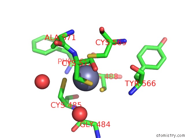
Mono view
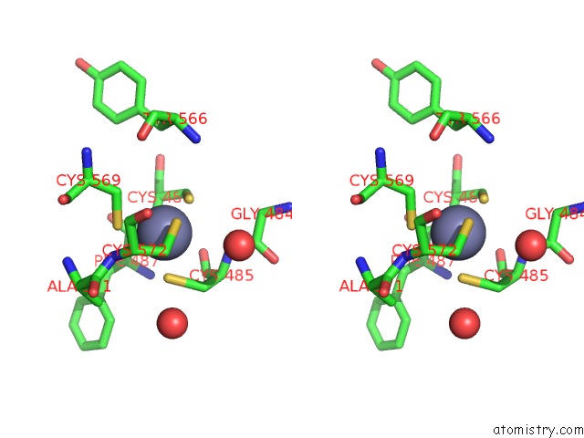
Stereo pair view
Mono view
Stereo pair view
A full contact list of Zinc with other atoms in the Zn binding
site number 1 of Crystal Structure of ATG7C-ATG8 Complex within 5.0Å range:
|
Reference:
S.B.Hong,
B.W.Kim,
K.E.Lee,
S.W.Kim,
H.Jeon,
J.Kim,
H.K.Song.
Insights Into Noncanonical E1 Enzyme Activation From the Structure of Autophagic E1 ATG7 with ATG8. Nat.Struct.Mol.Biol. V. 18 1323 2011.
ISSN: ISSN 1545-9993
PubMed: 22056771
DOI: 10.1038/NSMB.2165
Page generated: Sat Oct 26 15:06:27 2024
ISSN: ISSN 1545-9993
PubMed: 22056771
DOI: 10.1038/NSMB.2165
Last articles
Fe in 2YXOFe in 2YRS
Fe in 2YXC
Fe in 2YNM
Fe in 2YVJ
Fe in 2YP1
Fe in 2YU2
Fe in 2YU1
Fe in 2YQB
Fe in 2YOO