Zinc »
PDB 3r0d-3rja »
3rfr »
Zinc in PDB 3rfr: Crystal Structure of Particulate Methane Monooxygenase (Pmmo) From Methylocystis Sp. Strain M
Protein crystallography data
The structure of Crystal Structure of Particulate Methane Monooxygenase (Pmmo) From Methylocystis Sp. Strain M, PDB code: 3rfr
was solved by
S.M.Smith,
A.C.Rosenzweig,
with X-Ray Crystallography technique. A brief refinement statistics is given in the table below:
| Resolution Low / High (Å) | 45.79 / 2.68 |
| Space group | P 21 21 21 |
| Cell size a, b, c (Å), α, β, γ (°) | 107.718, 178.310, 183.147, 90.00, 90.00, 90.00 |
| R / Rfree (%) | 24.9 / 28.1 |
Other elements in 3rfr:
The structure of Crystal Structure of Particulate Methane Monooxygenase (Pmmo) From Methylocystis Sp. Strain M also contains other interesting chemical elements:
| Copper | (Cu) | 4 atoms |
Zinc Binding Sites:
The binding sites of Zinc atom in the Crystal Structure of Particulate Methane Monooxygenase (Pmmo) From Methylocystis Sp. Strain M
(pdb code 3rfr). This binding sites where shown within
5.0 Angstroms radius around Zinc atom.
In total 9 binding sites of Zinc where determined in the Crystal Structure of Particulate Methane Monooxygenase (Pmmo) From Methylocystis Sp. Strain M, PDB code: 3rfr:
Jump to Zinc binding site number: 1; 2; 3; 4; 5; 6; 7; 8; 9;
In total 9 binding sites of Zinc where determined in the Crystal Structure of Particulate Methane Monooxygenase (Pmmo) From Methylocystis Sp. Strain M, PDB code: 3rfr:
Jump to Zinc binding site number: 1; 2; 3; 4; 5; 6; 7; 8; 9;
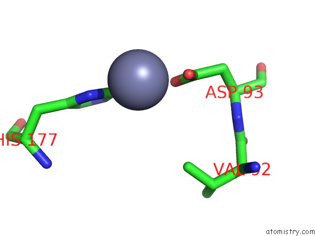
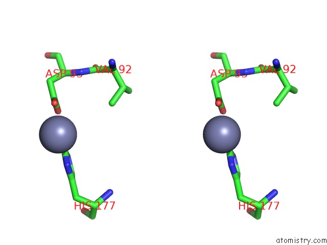
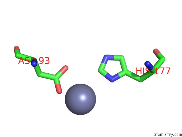
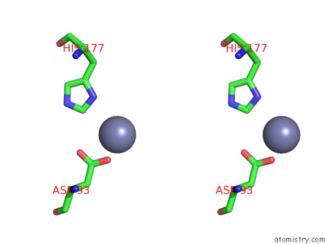
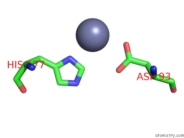
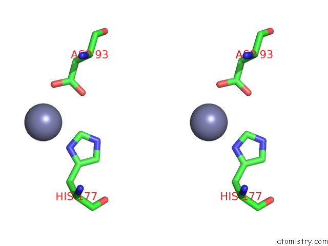
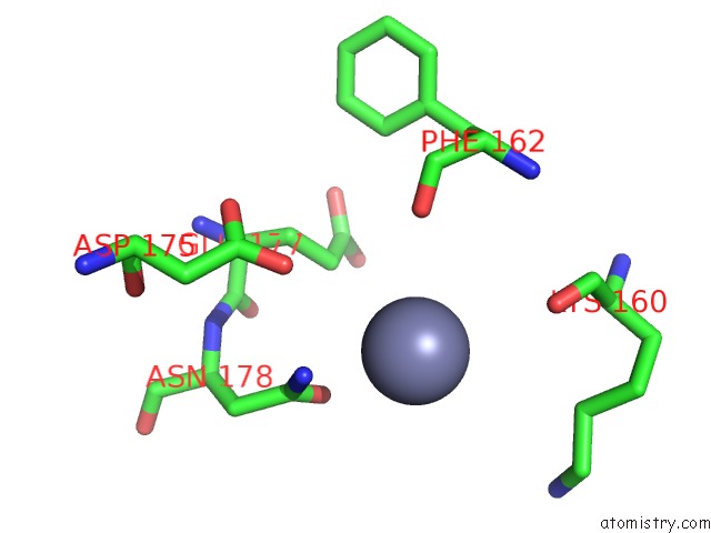
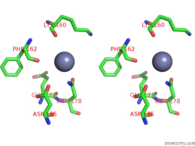
Zinc binding site 1 out of 9 in 3rfr
Go back to
Zinc binding site 1 out
of 9 in the Crystal Structure of Particulate Methane Monooxygenase (Pmmo) From Methylocystis Sp. Strain M
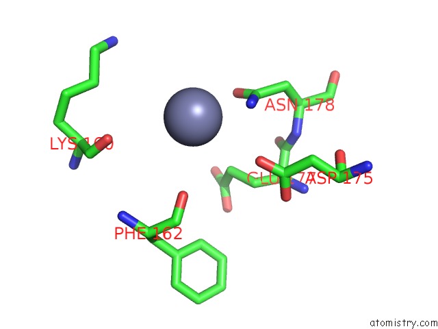
Mono view
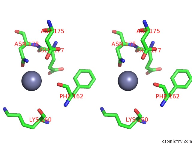
Stereo pair view
Mono view
Stereo pair view
A full contact list of Zinc with other atoms in the Zn binding
site number 1 of Crystal Structure of Particulate Methane Monooxygenase (Pmmo) From Methylocystis Sp. Strain M within 5.0Å range:
|
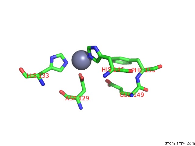
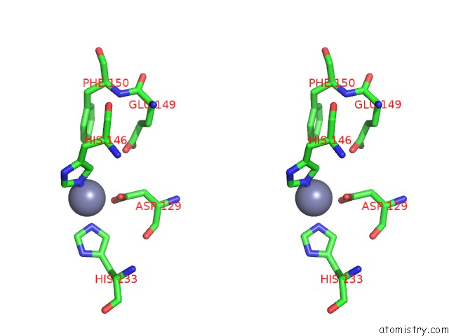
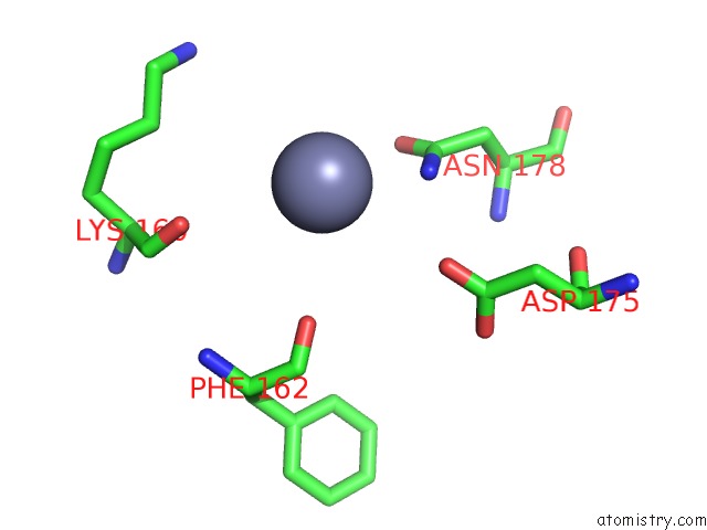
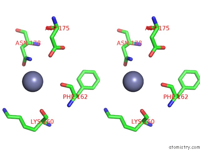
Zinc binding site 2 out of 9 in 3rfr
Go back to
Zinc binding site 2 out
of 9 in the Crystal Structure of Particulate Methane Monooxygenase (Pmmo) From Methylocystis Sp. Strain M
Mono view
Stereo pair view
Mono view
Stereo pair view
A full contact list of Zinc with other atoms in the Zn binding
site number 2 of Crystal Structure of Particulate Methane Monooxygenase (Pmmo) From Methylocystis Sp. Strain M within 5.0Å range:
|
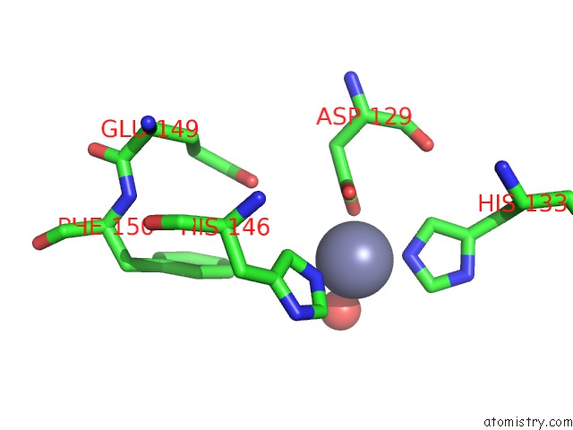
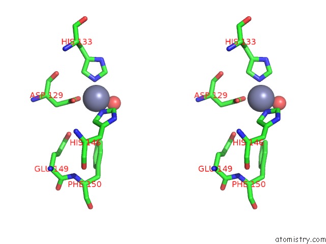
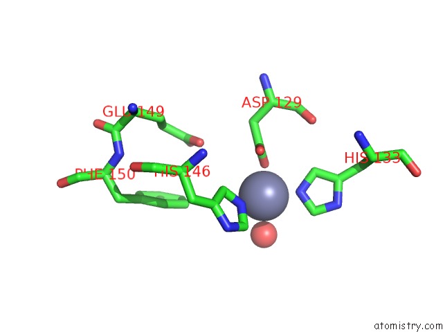
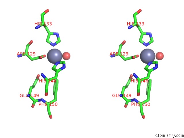
Zinc binding site 3 out of 9 in 3rfr
Go back to
Zinc binding site 3 out
of 9 in the Crystal Structure of Particulate Methane Monooxygenase (Pmmo) From Methylocystis Sp. Strain M
Mono view
Stereo pair view
Mono view
Stereo pair view
A full contact list of Zinc with other atoms in the Zn binding
site number 3 of Crystal Structure of Particulate Methane Monooxygenase (Pmmo) From Methylocystis Sp. Strain M within 5.0Å range:
|
Zinc binding site 4 out of 9 in 3rfr
Go back to
Zinc binding site 4 out
of 9 in the Crystal Structure of Particulate Methane Monooxygenase (Pmmo) From Methylocystis Sp. Strain M
Mono view
Stereo pair view
Mono view
Stereo pair view
A full contact list of Zinc with other atoms in the Zn binding
site number 4 of Crystal Structure of Particulate Methane Monooxygenase (Pmmo) From Methylocystis Sp. Strain M within 5.0Å range:
|
Zinc binding site 5 out of 9 in 3rfr
Go back to
Zinc binding site 5 out
of 9 in the Crystal Structure of Particulate Methane Monooxygenase (Pmmo) From Methylocystis Sp. Strain M
Mono view
Stereo pair view
Mono view
Stereo pair view
A full contact list of Zinc with other atoms in the Zn binding
site number 5 of Crystal Structure of Particulate Methane Monooxygenase (Pmmo) From Methylocystis Sp. Strain M within 5.0Å range:
|
Zinc binding site 6 out of 9 in 3rfr
Go back to
Zinc binding site 6 out
of 9 in the Crystal Structure of Particulate Methane Monooxygenase (Pmmo) From Methylocystis Sp. Strain M
Mono view
Stereo pair view
Mono view
Stereo pair view
A full contact list of Zinc with other atoms in the Zn binding
site number 6 of Crystal Structure of Particulate Methane Monooxygenase (Pmmo) From Methylocystis Sp. Strain M within 5.0Å range:
|
Zinc binding site 7 out of 9 in 3rfr
Go back to
Zinc binding site 7 out
of 9 in the Crystal Structure of Particulate Methane Monooxygenase (Pmmo) From Methylocystis Sp. Strain M
Mono view
Stereo pair view
Mono view
Stereo pair view
A full contact list of Zinc with other atoms in the Zn binding
site number 7 of Crystal Structure of Particulate Methane Monooxygenase (Pmmo) From Methylocystis Sp. Strain M within 5.0Å range:
|
Zinc binding site 8 out of 9 in 3rfr
Go back to
Zinc binding site 8 out
of 9 in the Crystal Structure of Particulate Methane Monooxygenase (Pmmo) From Methylocystis Sp. Strain M
Mono view
Stereo pair view
Mono view
Stereo pair view
A full contact list of Zinc with other atoms in the Zn binding
site number 8 of Crystal Structure of Particulate Methane Monooxygenase (Pmmo) From Methylocystis Sp. Strain M within 5.0Å range:
|
Zinc binding site 9 out of 9 in 3rfr
Go back to
Zinc binding site 9 out
of 9 in the Crystal Structure of Particulate Methane Monooxygenase (Pmmo) From Methylocystis Sp. Strain M
Mono view
Stereo pair view
Mono view
Stereo pair view
A full contact list of Zinc with other atoms in the Zn binding
site number 9 of Crystal Structure of Particulate Methane Monooxygenase (Pmmo) From Methylocystis Sp. Strain M within 5.0Å range:
|
Reference:
S.M.Smith,
S.Rawat,
J.Telser,
B.M.Hoffman,
T.L.Stemmler,
A.C.Rosenzweig.
Crystal Structure and Characterization of Particulate Methane Monooxygenase From Methylocystis Species Strain M. Biochemistry V. 50 10231 2011.
ISSN: ISSN 0006-2960
PubMed: 22013879
DOI: 10.1021/BI200801Z
Page generated: Sat Oct 26 14:45:37 2024
ISSN: ISSN 0006-2960
PubMed: 22013879
DOI: 10.1021/BI200801Z
Last articles
I in 1HK4I in 1GW9
I in 1GWA
I in 1HDY
I in 1HC0
I in 1HC9
I in 1GWG
I in 1GZA
I in 1GWD
I in 1GUL