Zinc »
PDB 3phx-3ptm »
3pnu »
Zinc in PDB 3pnu: 2.4 Angstrom Crystal Structure of Dihydroorotase (Pyrc) From Campylobacter Jejuni.
Enzymatic activity of 2.4 Angstrom Crystal Structure of Dihydroorotase (Pyrc) From Campylobacter Jejuni.
All present enzymatic activity of 2.4 Angstrom Crystal Structure of Dihydroorotase (Pyrc) From Campylobacter Jejuni.:
3.5.2.3;
3.5.2.3;
Protein crystallography data
The structure of 2.4 Angstrom Crystal Structure of Dihydroorotase (Pyrc) From Campylobacter Jejuni., PDB code: 3pnu
was solved by
G.Minasov,
A.Halavaty,
L.Shuvalova,
I.Dubrovska,
J.Winsor,
L.Papazisi,
W.F.Anderson,
Center For Structural Genomics Of Infectious Diseases(Csgid),
with X-Ray Crystallography technique. A brief refinement statistics is given in the table below:
| Resolution Low / High (Å) | 29.52 / 2.40 |
| Space group | P 21 21 21 |
| Cell size a, b, c (Å), α, β, γ (°) | 69.520, 80.802, 154.878, 90.00, 90.00, 90.00 |
| R / Rfree (%) | 19.1 / 25 |
Zinc Binding Sites:
The binding sites of Zinc atom in the 2.4 Angstrom Crystal Structure of Dihydroorotase (Pyrc) From Campylobacter Jejuni.
(pdb code 3pnu). This binding sites where shown within
5.0 Angstroms radius around Zinc atom.
In total 4 binding sites of Zinc where determined in the 2.4 Angstrom Crystal Structure of Dihydroorotase (Pyrc) From Campylobacter Jejuni., PDB code: 3pnu:
Jump to Zinc binding site number: 1; 2; 3; 4;
In total 4 binding sites of Zinc where determined in the 2.4 Angstrom Crystal Structure of Dihydroorotase (Pyrc) From Campylobacter Jejuni., PDB code: 3pnu:
Jump to Zinc binding site number: 1; 2; 3; 4;
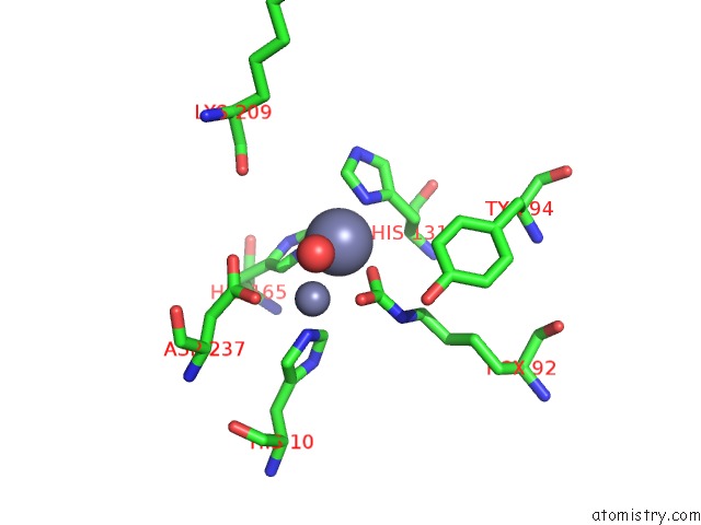
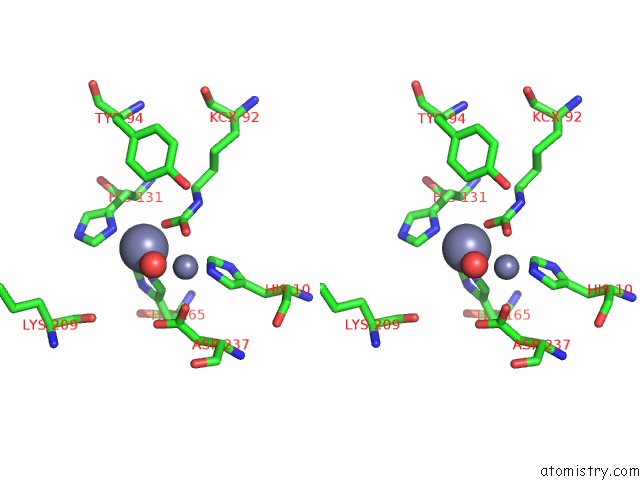
Zinc binding site 1 out of 4 in 3pnu
Go back to
Zinc binding site 1 out
of 4 in the 2.4 Angstrom Crystal Structure of Dihydroorotase (Pyrc) From Campylobacter Jejuni.
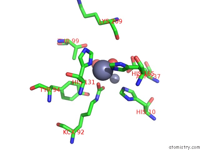
Mono view
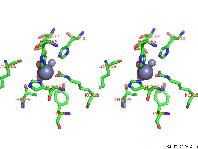
Stereo pair view
Mono view
Stereo pair view
A full contact list of Zinc with other atoms in the Zn binding
site number 1 of 2.4 Angstrom Crystal Structure of Dihydroorotase (Pyrc) From Campylobacter Jejuni. within 5.0Å range:
|
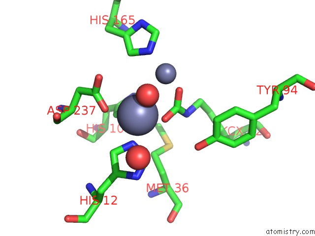
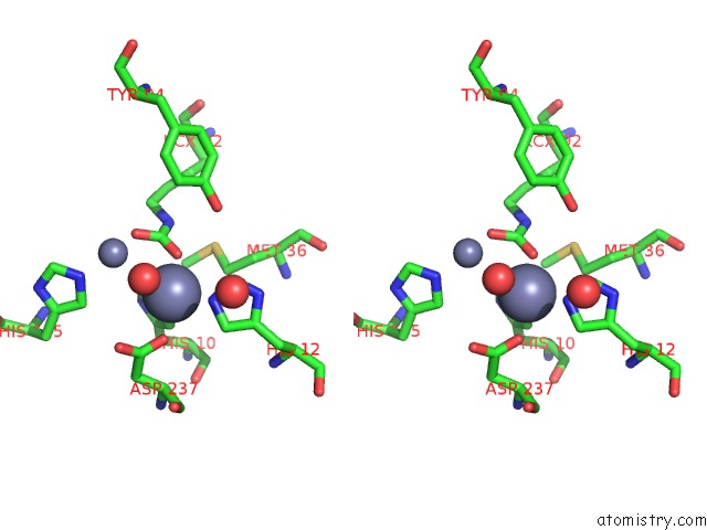
Zinc binding site 2 out of 4 in 3pnu
Go back to
Zinc binding site 2 out
of 4 in the 2.4 Angstrom Crystal Structure of Dihydroorotase (Pyrc) From Campylobacter Jejuni.
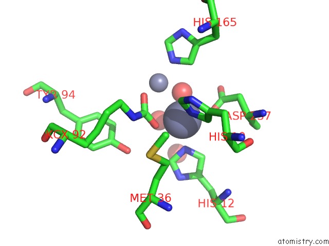
Mono view
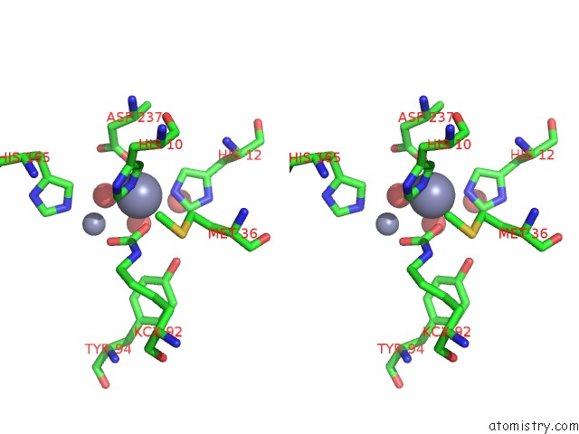
Stereo pair view
Mono view
Stereo pair view
A full contact list of Zinc with other atoms in the Zn binding
site number 2 of 2.4 Angstrom Crystal Structure of Dihydroorotase (Pyrc) From Campylobacter Jejuni. within 5.0Å range:
|
Zinc binding site 3 out of 4 in 3pnu
Go back to
Zinc binding site 3 out
of 4 in the 2.4 Angstrom Crystal Structure of Dihydroorotase (Pyrc) From Campylobacter Jejuni.
Mono view
Stereo pair view
Mono view
Stereo pair view
A full contact list of Zinc with other atoms in the Zn binding
site number 3 of 2.4 Angstrom Crystal Structure of Dihydroorotase (Pyrc) From Campylobacter Jejuni. within 5.0Å range:
|
Zinc binding site 4 out of 4 in 3pnu
Go back to
Zinc binding site 4 out
of 4 in the 2.4 Angstrom Crystal Structure of Dihydroorotase (Pyrc) From Campylobacter Jejuni.
Mono view
Stereo pair view
Mono view
Stereo pair view
A full contact list of Zinc with other atoms in the Zn binding
site number 4 of 2.4 Angstrom Crystal Structure of Dihydroorotase (Pyrc) From Campylobacter Jejuni. within 5.0Å range:
|
Reference:
G.Minasov,
A.Halavaty,
L.Shuvalova,
I.Dubrovska,
J.Winsor,
L.Papazisi,
W.F.Anderson,
Center For Structural Genomics Of Infectious Diseases(Csgid).
2.4 Angstrom Crystal Structure of Dihydroorotase (Pyrc) From Campylobacter Jejuni. To Be Published.
Page generated: Sat Oct 26 11:37:12 2024
Last articles
Mg in 6EM6Mg in 6ELI
Mg in 6EL4
Mg in 6EL0
Mg in 6EKZ
Mg in 6EKY
Mg in 6EKX
Mg in 6EHS
Mg in 6EKW
Mg in 6EKH