Zinc »
PDB 3o7a-3okk »
3ofi »
Zinc in PDB 3ofi: Crystal Structure of Human Insulin-Degrading Enzyme in Complex with Ubiquitin
Enzymatic activity of Crystal Structure of Human Insulin-Degrading Enzyme in Complex with Ubiquitin
All present enzymatic activity of Crystal Structure of Human Insulin-Degrading Enzyme in Complex with Ubiquitin:
3.4.24.56;
3.4.24.56;
Protein crystallography data
The structure of Crystal Structure of Human Insulin-Degrading Enzyme in Complex with Ubiquitin, PDB code: 3ofi
was solved by
V.Kalas,
L.A.Ralat,
W.-J.Tang,
with X-Ray Crystallography technique. A brief refinement statistics is given in the table below:
| Resolution Low / High (Å) | 50.00 / 2.35 |
| Space group | P 65 |
| Cell size a, b, c (Å), α, β, γ (°) | 262.943, 262.943, 90.968, 90.00, 90.00, 120.00 |
| R / Rfree (%) | 21 / 24 |
Zinc Binding Sites:
The binding sites of Zinc atom in the Crystal Structure of Human Insulin-Degrading Enzyme in Complex with Ubiquitin
(pdb code 3ofi). This binding sites where shown within
5.0 Angstroms radius around Zinc atom.
In total 2 binding sites of Zinc where determined in the Crystal Structure of Human Insulin-Degrading Enzyme in Complex with Ubiquitin, PDB code: 3ofi:
Jump to Zinc binding site number: 1; 2;
In total 2 binding sites of Zinc where determined in the Crystal Structure of Human Insulin-Degrading Enzyme in Complex with Ubiquitin, PDB code: 3ofi:
Jump to Zinc binding site number: 1; 2;
Zinc binding site 1 out of 2 in 3ofi
Go back to
Zinc binding site 1 out
of 2 in the Crystal Structure of Human Insulin-Degrading Enzyme in Complex with Ubiquitin
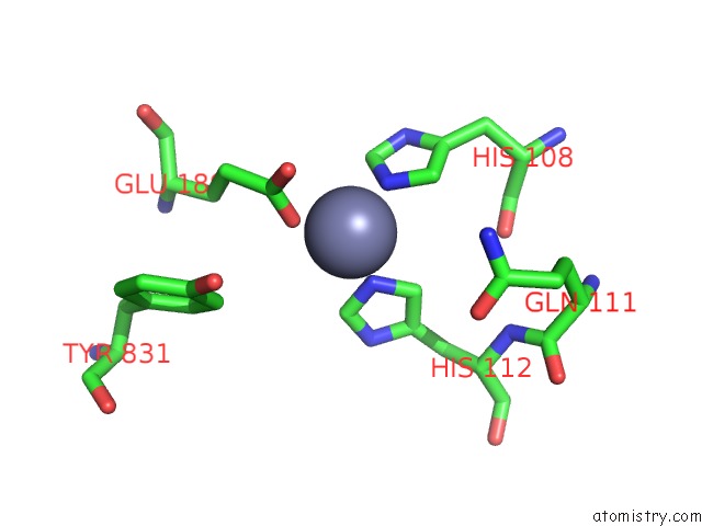
Mono view
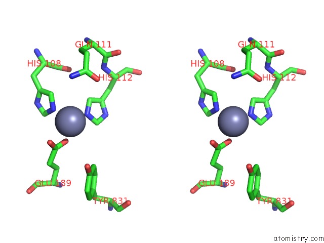
Stereo pair view
Mono view
Stereo pair view
A full contact list of Zinc with other atoms in the Zn binding
site number 1 of Crystal Structure of Human Insulin-Degrading Enzyme in Complex with Ubiquitin within 5.0Å range:
|
Zinc binding site 2 out of 2 in 3ofi
Go back to
Zinc binding site 2 out
of 2 in the Crystal Structure of Human Insulin-Degrading Enzyme in Complex with Ubiquitin
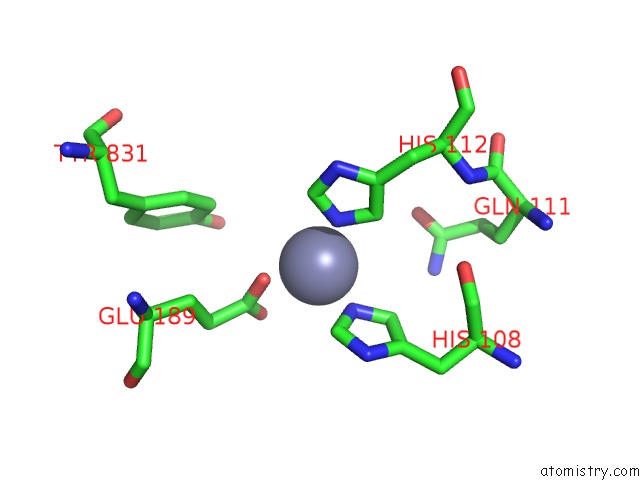
Mono view
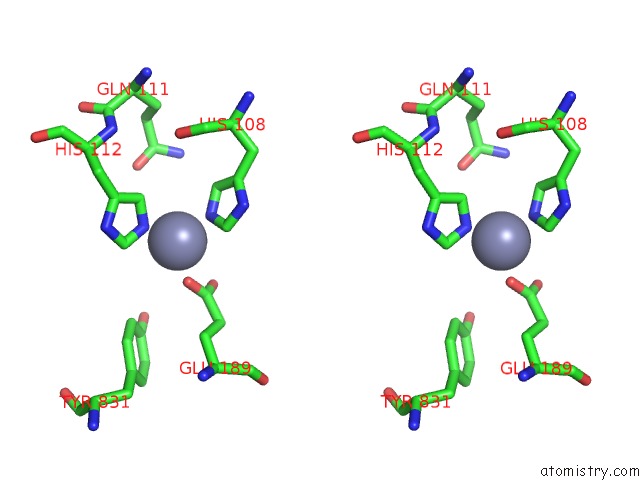
Stereo pair view
Mono view
Stereo pair view
A full contact list of Zinc with other atoms in the Zn binding
site number 2 of Crystal Structure of Human Insulin-Degrading Enzyme in Complex with Ubiquitin within 5.0Å range:
|
Reference:
L.A.Ralat,
V.Kalas,
Z.Zheng,
R.D.Goldman,
T.R.Sosnick,
W.J.Tang.
Ubiquitin Is A Novel Substrate For Human Insulin-Degrading Enzyme. J.Mol.Biol. V. 406 454 2011.
ISSN: ISSN 0022-2836
PubMed: 21185309
DOI: 10.1016/J.JMB.2010.12.026
Page generated: Sat Oct 26 10:55:06 2024
ISSN: ISSN 0022-2836
PubMed: 21185309
DOI: 10.1016/J.JMB.2010.12.026
Last articles
Fe in 2YXOFe in 2YRS
Fe in 2YXC
Fe in 2YNM
Fe in 2YVJ
Fe in 2YP1
Fe in 2YU2
Fe in 2YU1
Fe in 2YQB
Fe in 2YOO