Zinc »
PDB 3m6r-3mhx »
3mdj »
Zinc in PDB 3mdj: Er Aminopeptidase, ERAP1, Bound to the Zinc Aminopeptidase Inhibitor, Bestatin
Protein crystallography data
The structure of Er Aminopeptidase, ERAP1, Bound to the Zinc Aminopeptidase Inhibitor, Bestatin, PDB code: 3mdj
was solved by
T.T.Nguyen,
L.J.Stern,
with X-Ray Crystallography technique. A brief refinement statistics is given in the table below:
| Resolution Low / High (Å) | 38.11 / 2.95 |
| Space group | P 1 21 1 |
| Cell size a, b, c (Å), α, β, γ (°) | 71.029, 234.635, 95.860, 90.00, 103.59, 90.00 |
| R / Rfree (%) | 19.9 / 26.4 |
Zinc Binding Sites:
The binding sites of Zinc atom in the Er Aminopeptidase, ERAP1, Bound to the Zinc Aminopeptidase Inhibitor, Bestatin
(pdb code 3mdj). This binding sites where shown within
5.0 Angstroms radius around Zinc atom.
In total 3 binding sites of Zinc where determined in the Er Aminopeptidase, ERAP1, Bound to the Zinc Aminopeptidase Inhibitor, Bestatin, PDB code: 3mdj:
Jump to Zinc binding site number: 1; 2; 3;
In total 3 binding sites of Zinc where determined in the Er Aminopeptidase, ERAP1, Bound to the Zinc Aminopeptidase Inhibitor, Bestatin, PDB code: 3mdj:
Jump to Zinc binding site number: 1; 2; 3;
Zinc binding site 1 out of 3 in 3mdj
Go back to Zinc Binding Sites List in 3mdj
Zinc binding site 1 out
of 3 in the Er Aminopeptidase, ERAP1, Bound to the Zinc Aminopeptidase Inhibitor, Bestatin
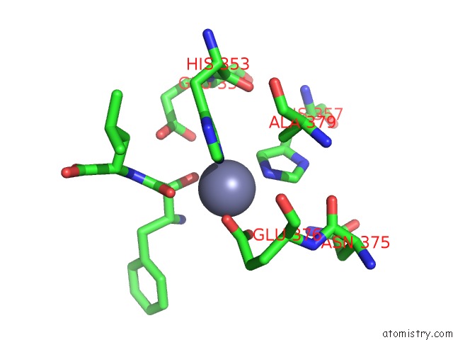
Mono view
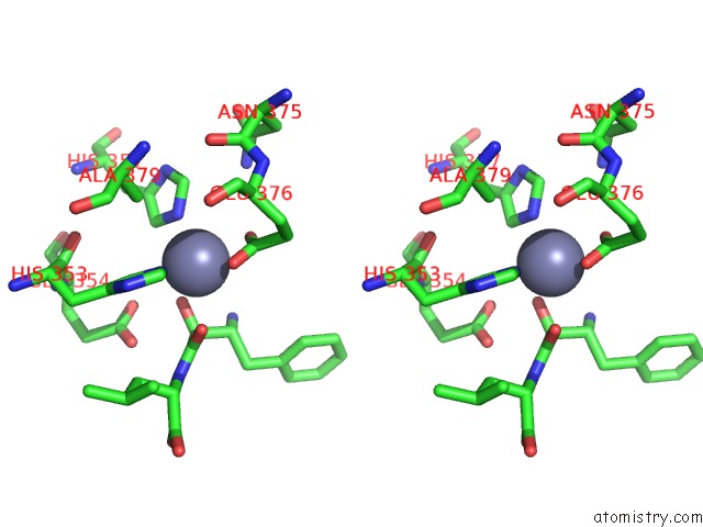
Stereo pair view
Mono view
Stereo pair view
A full contact list of Zinc with other atoms in the Zn binding
site number 1 of Er Aminopeptidase, ERAP1, Bound to the Zinc Aminopeptidase Inhibitor, Bestatin within 5.0Å range:
|
Zinc binding site 2 out of 3 in 3mdj
Go back to
Zinc Binding Sites List in 3mdj
Zinc binding site 2 out
of 3 in the Er Aminopeptidase, ERAP1, Bound to the Zinc Aminopeptidase Inhibitor, Bestatin
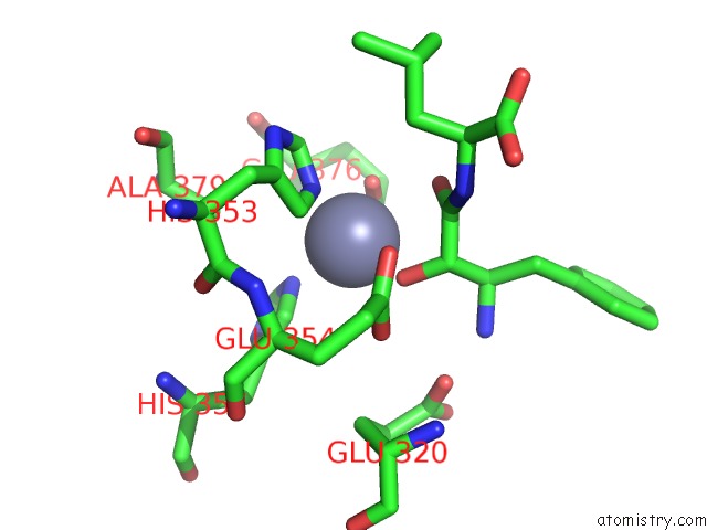
Mono view
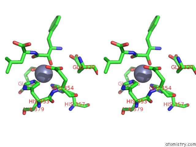
Stereo pair view
Mono view
Stereo pair view
A full contact list of Zinc with other atoms in the Zn binding
site number 2 of Er Aminopeptidase, ERAP1, Bound to the Zinc Aminopeptidase Inhibitor, Bestatin within 5.0Å range:
|
Zinc binding site 3 out of 3 in 3mdj
Go back to
Zinc Binding Sites List in 3mdj
Zinc binding site 3 out
of 3 in the Er Aminopeptidase, ERAP1, Bound to the Zinc Aminopeptidase Inhibitor, Bestatin
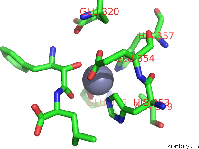
Mono view
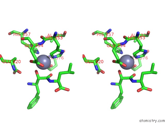
Stereo pair view
Mono view
Stereo pair view
A full contact list of Zinc with other atoms in the Zn binding
site number 3 of Er Aminopeptidase, ERAP1, Bound to the Zinc Aminopeptidase Inhibitor, Bestatin within 5.0Å range:
|
Reference:
T.T.Nguyen,
S.C.Chang,
I.Evnouchidou,
I.A.York,
C.Zikos,
K.L.Rock,
A.L.Goldberg,
E.Stratikos,
L.J.Stern.
Structural Basis For Antigenic Peptide Precursor Processing By the Endoplasmic Reticulum Aminopeptidase ERAP1. Nat.Struct.Mol.Biol. V. 18 604 2011.
ISSN: ISSN 1545-9993
PubMed: 21478864
DOI: 10.1038/NSMB.2021
Page generated: Sat Oct 26 09:25:01 2024
ISSN: ISSN 1545-9993
PubMed: 21478864
DOI: 10.1038/NSMB.2021
Last articles
Fe in 7R0FFe in 7QZH
Fe in 7QZR
Fe in 7QZG
Fe in 7QZF
Fe in 7QZA
Fe in 7QZE
Fe in 7QSO
Fe in 7QYZ
Fe in 7QYQ
