Zinc »
PDB 3m6r-3mhx »
3mbj »
Zinc in PDB 3mbj: Crystal Structure of A Putative Phosphomethylpyrimidine Kinase (BT_4458) From Bacteroides Thetaiotaomicron Vpi-5482 at 2.10 A Resolution (Rhombohedral Form)
Protein crystallography data
The structure of Crystal Structure of A Putative Phosphomethylpyrimidine Kinase (BT_4458) From Bacteroides Thetaiotaomicron Vpi-5482 at 2.10 A Resolution (Rhombohedral Form), PDB code: 3mbj
was solved by
Joint Center For Structural Genomics (Jcsg),
with X-Ray Crystallography technique. A brief refinement statistics is given in the table below:
| Resolution Low / High (Å) | 29.59 / 2.10 |
| Space group | H 3 2 |
| Cell size a, b, c (Å), α, β, γ (°) | 161.627, 161.627, 76.138, 90.00, 90.00, 120.00 |
| R / Rfree (%) | 17.1 / 21 |
Other elements in 3mbj:
The structure of Crystal Structure of A Putative Phosphomethylpyrimidine Kinase (BT_4458) From Bacteroides Thetaiotaomicron Vpi-5482 at 2.10 A Resolution (Rhombohedral Form) also contains other interesting chemical elements:
| Chlorine | (Cl) | 1 atom |
Zinc Binding Sites:
The binding sites of Zinc atom in the Crystal Structure of A Putative Phosphomethylpyrimidine Kinase (BT_4458) From Bacteroides Thetaiotaomicron Vpi-5482 at 2.10 A Resolution (Rhombohedral Form)
(pdb code 3mbj). This binding sites where shown within
5.0 Angstroms radius around Zinc atom.
In total 3 binding sites of Zinc where determined in the Crystal Structure of A Putative Phosphomethylpyrimidine Kinase (BT_4458) From Bacteroides Thetaiotaomicron Vpi-5482 at 2.10 A Resolution (Rhombohedral Form), PDB code: 3mbj:
Jump to Zinc binding site number: 1; 2; 3;
In total 3 binding sites of Zinc where determined in the Crystal Structure of A Putative Phosphomethylpyrimidine Kinase (BT_4458) From Bacteroides Thetaiotaomicron Vpi-5482 at 2.10 A Resolution (Rhombohedral Form), PDB code: 3mbj:
Jump to Zinc binding site number: 1; 2; 3;
Zinc binding site 1 out of 3 in 3mbj
Go back to Zinc Binding Sites List in 3mbj
Zinc binding site 1 out
of 3 in the Crystal Structure of A Putative Phosphomethylpyrimidine Kinase (BT_4458) From Bacteroides Thetaiotaomicron Vpi-5482 at 2.10 A Resolution (Rhombohedral Form)
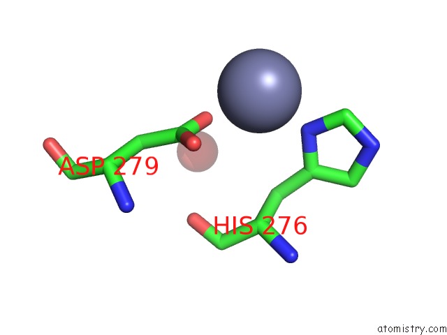
Mono view
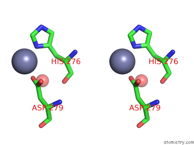
Stereo pair view
Mono view
Stereo pair view
A full contact list of Zinc with other atoms in the Zn binding
site number 1 of Crystal Structure of A Putative Phosphomethylpyrimidine Kinase (BT_4458) From Bacteroides Thetaiotaomicron Vpi-5482 at 2.10 A Resolution (Rhombohedral Form) within 5.0Å range:
|
Zinc binding site 2 out of 3 in 3mbj
Go back to
Zinc Binding Sites List in 3mbj
Zinc binding site 2 out
of 3 in the Crystal Structure of A Putative Phosphomethylpyrimidine Kinase (BT_4458) From Bacteroides Thetaiotaomicron Vpi-5482 at 2.10 A Resolution (Rhombohedral Form)
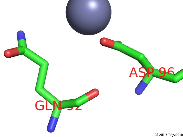
Mono view
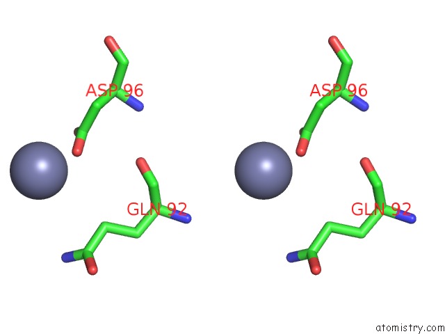
Stereo pair view
Mono view
Stereo pair view
A full contact list of Zinc with other atoms in the Zn binding
site number 2 of Crystal Structure of A Putative Phosphomethylpyrimidine Kinase (BT_4458) From Bacteroides Thetaiotaomicron Vpi-5482 at 2.10 A Resolution (Rhombohedral Form) within 5.0Å range:
|
Zinc binding site 3 out of 3 in 3mbj
Go back to
Zinc Binding Sites List in 3mbj
Zinc binding site 3 out
of 3 in the Crystal Structure of A Putative Phosphomethylpyrimidine Kinase (BT_4458) From Bacteroides Thetaiotaomicron Vpi-5482 at 2.10 A Resolution (Rhombohedral Form)
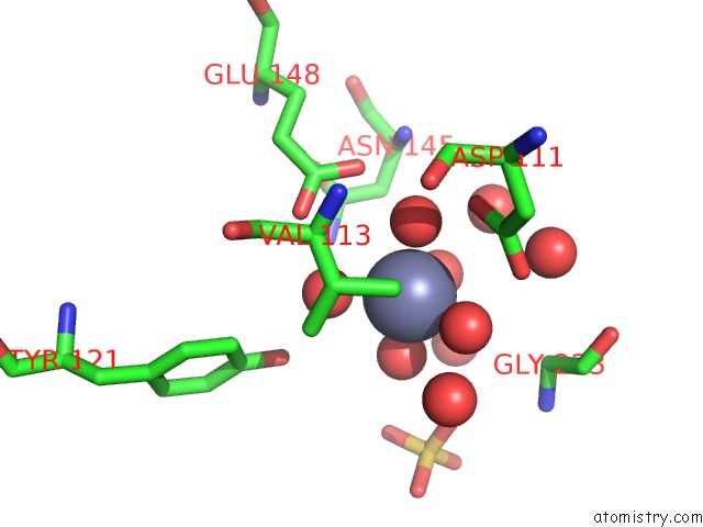
Mono view
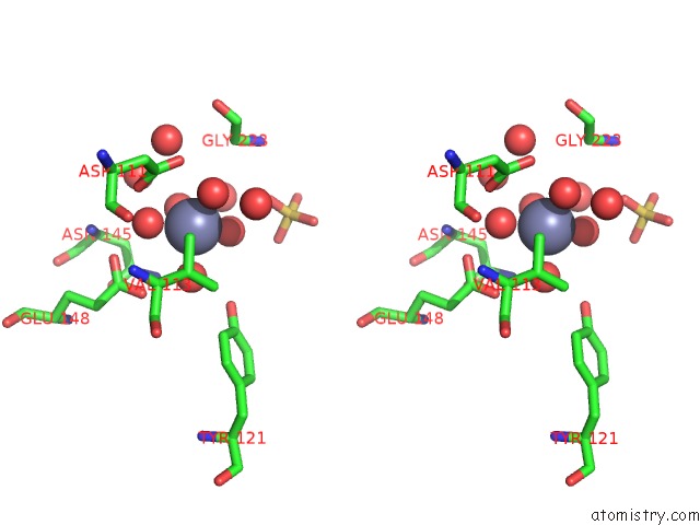
Stereo pair view
Mono view
Stereo pair view
A full contact list of Zinc with other atoms in the Zn binding
site number 3 of Crystal Structure of A Putative Phosphomethylpyrimidine Kinase (BT_4458) From Bacteroides Thetaiotaomicron Vpi-5482 at 2.10 A Resolution (Rhombohedral Form) within 5.0Å range:
|
Reference:
Joint Center For Structural Genomics (Jcsg),
Joint Center For Structural Genomics (Jcsg).
N/A N/A.
Page generated: Sat Oct 26 09:22:45 2024
Last articles
Fe in 7QZGFe in 7QZF
Fe in 7QZA
Fe in 7QZE
Fe in 7QSO
Fe in 7QYZ
Fe in 7QYQ
Fe in 7QSN
Fe in 7QWN
Fe in 7QTH
