Zinc »
PDB 3lta-3m1m »
3ltd »
Zinc in PDB 3ltd: X-Ray Structure of A Non-Biological Atp Binding Protein Determined at 2.8 A By Multi-Wavelength Anomalous Dispersion
Protein crystallography data
The structure of X-Ray Structure of A Non-Biological Atp Binding Protein Determined at 2.8 A By Multi-Wavelength Anomalous Dispersion, PDB code: 3ltd
was solved by
C.R.Simmons,
C.L.Magee,
J.P.Allen,
J.C.Chaput,
with X-Ray Crystallography technique. A brief refinement statistics is given in the table below:
| Resolution Low / High (Å) | 36.56 / 2.80 |
| Space group | P 32 2 1 |
| Cell size a, b, c (Å), α, β, γ (°) | 73.145, 73.145, 54.804, 90.00, 90.00, 120.00 |
| R / Rfree (%) | 18 / 21.8 |
Other elements in 3ltd:
The structure of X-Ray Structure of A Non-Biological Atp Binding Protein Determined at 2.8 A By Multi-Wavelength Anomalous Dispersion also contains other interesting chemical elements:
| Chlorine | (Cl) | 1 atom |
Zinc Binding Sites:
The binding sites of Zinc atom in the X-Ray Structure of A Non-Biological Atp Binding Protein Determined at 2.8 A By Multi-Wavelength Anomalous Dispersion
(pdb code 3ltd). This binding sites where shown within
5.0 Angstroms radius around Zinc atom.
In total only one binding site of Zinc was determined in the X-Ray Structure of A Non-Biological Atp Binding Protein Determined at 2.8 A By Multi-Wavelength Anomalous Dispersion, PDB code: 3ltd:
In total only one binding site of Zinc was determined in the X-Ray Structure of A Non-Biological Atp Binding Protein Determined at 2.8 A By Multi-Wavelength Anomalous Dispersion, PDB code: 3ltd:
Zinc binding site 1 out of 1 in 3ltd
Go back to
Zinc binding site 1 out
of 1 in the X-Ray Structure of A Non-Biological Atp Binding Protein Determined at 2.8 A By Multi-Wavelength Anomalous Dispersion
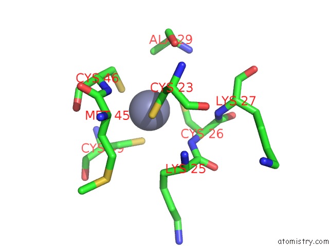
Mono view
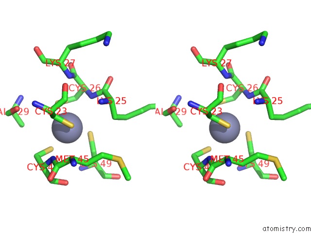
Stereo pair view
Mono view
Stereo pair view
A full contact list of Zinc with other atoms in the Zn binding
site number 1 of X-Ray Structure of A Non-Biological Atp Binding Protein Determined at 2.8 A By Multi-Wavelength Anomalous Dispersion within 5.0Å range:
|
Reference:
C.R.Simmons,
C.L.Magee,
D.A.Smith,
L.Lauman,
J.C.Chaput,
J.P.Allen.
Three-Dimensional Structures Reveal Multiple Adp/Atp Binding Modes For A Synthetic Class of Artificial Proteins. Biochemistry V. 49 8689 2010.
ISSN: ISSN 0006-2960
PubMed: 20822107
DOI: 10.1021/BI100398P
Page generated: Sat Oct 26 08:52:59 2024
ISSN: ISSN 0006-2960
PubMed: 20822107
DOI: 10.1021/BI100398P
Last articles
K in 6Z7VK in 6ZK9
K in 6Z8X
K in 6Z8W
K in 6Z8V
K in 6Z2K
K in 6Z2J
K in 6Z16
K in 6Z2V
K in 6Z0C