Zinc »
PDB 3lju-3lt9 »
3ls1 »
Zinc in PDB 3ls1: Crystal Structure of Cyanobacterial Psbq From Synechocystis Sp. Pcc 6803 Complexed with ZN2+
Protein crystallography data
The structure of Crystal Structure of Cyanobacterial Psbq From Synechocystis Sp. Pcc 6803 Complexed with ZN2+, PDB code: 3ls1
was solved by
S.A.Jackson,
R.D.Fagerlund,
S.M.Wilbanks,
J.J.Eaton-Rye,
with X-Ray Crystallography technique. A brief refinement statistics is given in the table below:
| Resolution Low / High (Å) | 21.76 / 1.85 |
| Space group | I 1 2 1 |
| Cell size a, b, c (Å), α, β, γ (°) | 92.771, 29.030, 99.503, 90.00, 97.77, 90.00 |
| R / Rfree (%) | 17.9 / 21.8 |
Zinc Binding Sites:
The binding sites of Zinc atom in the Crystal Structure of Cyanobacterial Psbq From Synechocystis Sp. Pcc 6803 Complexed with ZN2+
(pdb code 3ls1). This binding sites where shown within
5.0 Angstroms radius around Zinc atom.
In total 4 binding sites of Zinc where determined in the Crystal Structure of Cyanobacterial Psbq From Synechocystis Sp. Pcc 6803 Complexed with ZN2+, PDB code: 3ls1:
Jump to Zinc binding site number: 1; 2; 3; 4;
In total 4 binding sites of Zinc where determined in the Crystal Structure of Cyanobacterial Psbq From Synechocystis Sp. Pcc 6803 Complexed with ZN2+, PDB code: 3ls1:
Jump to Zinc binding site number: 1; 2; 3; 4;
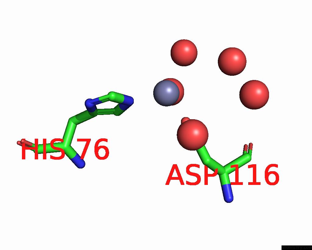
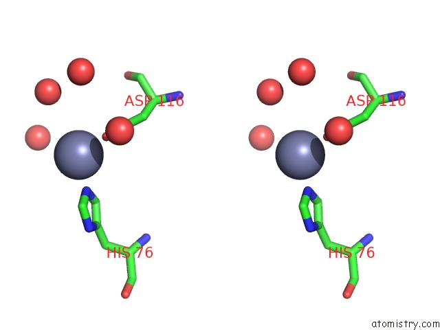
Zinc binding site 1 out of 4 in 3ls1
Go back to
Zinc binding site 1 out
of 4 in the Crystal Structure of Cyanobacterial Psbq From Synechocystis Sp. Pcc 6803 Complexed with ZN2+
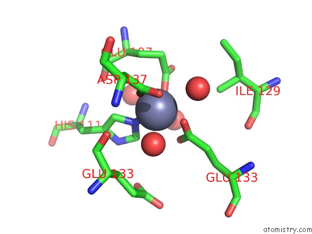
Mono view
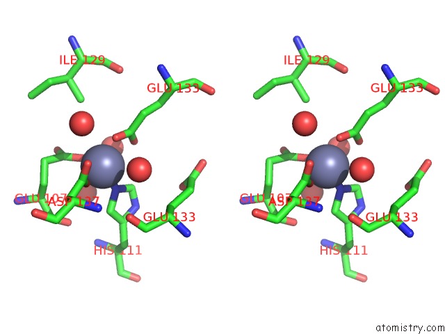
Stereo pair view
Mono view
Stereo pair view
A full contact list of Zinc with other atoms in the Zn binding
site number 1 of Crystal Structure of Cyanobacterial Psbq From Synechocystis Sp. Pcc 6803 Complexed with ZN2+ within 5.0Å range:
|
Zinc binding site 2 out of 4 in 3ls1
Go back to
Zinc binding site 2 out
of 4 in the Crystal Structure of Cyanobacterial Psbq From Synechocystis Sp. Pcc 6803 Complexed with ZN2+
Mono view
Stereo pair view
Mono view
Stereo pair view
A full contact list of Zinc with other atoms in the Zn binding
site number 2 of Crystal Structure of Cyanobacterial Psbq From Synechocystis Sp. Pcc 6803 Complexed with ZN2+ within 5.0Å range:
|
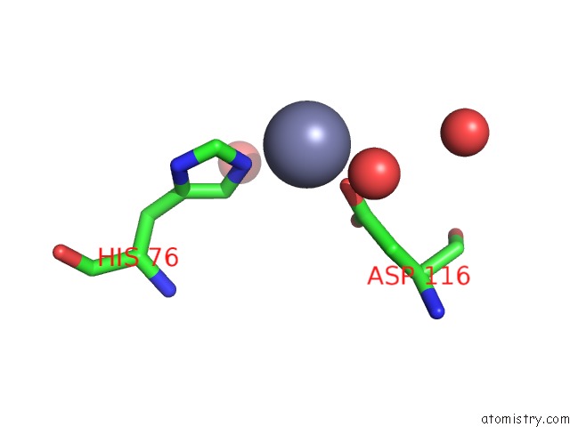
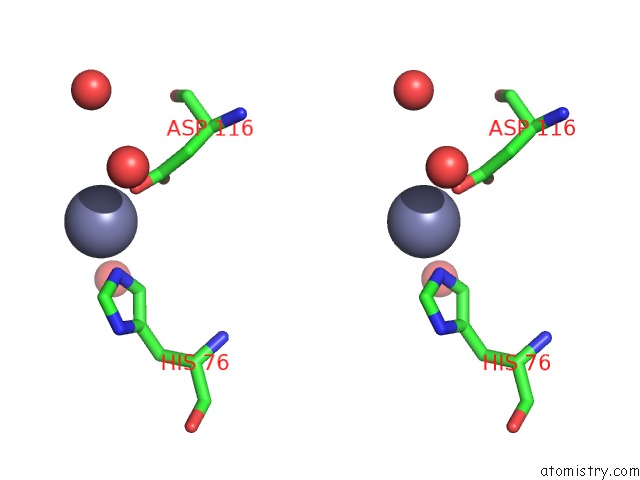
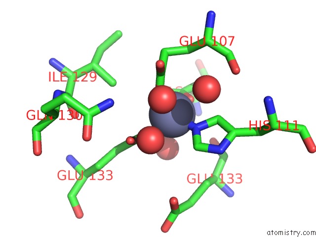
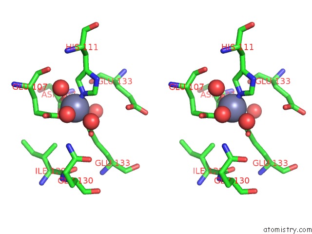
Zinc binding site 3 out of 4 in 3ls1
Go back to
Zinc binding site 3 out
of 4 in the Crystal Structure of Cyanobacterial Psbq From Synechocystis Sp. Pcc 6803 Complexed with ZN2+
Mono view
Stereo pair view
Mono view
Stereo pair view
A full contact list of Zinc with other atoms in the Zn binding
site number 3 of Crystal Structure of Cyanobacterial Psbq From Synechocystis Sp. Pcc 6803 Complexed with ZN2+ within 5.0Å range:
|
Zinc binding site 4 out of 4 in 3ls1
Go back to
Zinc binding site 4 out
of 4 in the Crystal Structure of Cyanobacterial Psbq From Synechocystis Sp. Pcc 6803 Complexed with ZN2+
Mono view
Stereo pair view
Mono view
Stereo pair view
A full contact list of Zinc with other atoms in the Zn binding
site number 4 of Crystal Structure of Cyanobacterial Psbq From Synechocystis Sp. Pcc 6803 Complexed with ZN2+ within 5.0Å range:
|
Reference:
S.A.Jackson,
R.D.Fagerlund,
S.M.Wilbanks,
J.J.Eaton-Rye.
Crystal Structure of Psbq From Synechocystis Sp. Pcc 6803 at 1.8 A: Implications For Binding and Function in Cyanobacterial Photosystem II Biochemistry V. 49 2765 2010.
ISSN: ISSN 0006-2960
PubMed: 20210304
DOI: 10.1021/BI100217H
Page generated: Sat Oct 26 08:48:13 2024
ISSN: ISSN 0006-2960
PubMed: 20210304
DOI: 10.1021/BI100217H
Last articles
K in 9ED0K in 9GEF
K in 9GBY
K in 9G9V
K in 9G9X
K in 9G9W
K in 9FDA
K in 9FCO
K in 9G5E
K in 9G5D