Zinc »
PDB 3k14-3kek »
3kde »
Zinc in PDB 3kde: Crystal Structure of the Thap Domain From D. Melanogaster P-Element Transposase in Complex with Its Natural Dna Binding Site
Protein crystallography data
The structure of Crystal Structure of the Thap Domain From D. Melanogaster P-Element Transposase in Complex with Its Natural Dna Binding Site, PDB code: 3kde
was solved by
A.Sabogal,
A.Y.Lyubimov,
J.M.Berger,
D.C.Rio,
with X-Ray Crystallography technique. A brief refinement statistics is given in the table below:
| Resolution Low / High (Å) | 35.09 / 1.74 |
| Space group | P 1 21 1 |
| Cell size a, b, c (Å), α, β, γ (°) | 28.692, 69.325, 35.126, 90.00, 92.52, 90.00 |
| R / Rfree (%) | 17.7 / 21.6 |
Other elements in 3kde:
The structure of Crystal Structure of the Thap Domain From D. Melanogaster P-Element Transposase in Complex with Its Natural Dna Binding Site also contains other interesting chemical elements:
| Bromine | (Br) | 2 atoms |
Zinc Binding Sites:
The binding sites of Zinc atom in the Crystal Structure of the Thap Domain From D. Melanogaster P-Element Transposase in Complex with Its Natural Dna Binding Site
(pdb code 3kde). This binding sites where shown within
5.0 Angstroms radius around Zinc atom.
In total only one binding site of Zinc was determined in the Crystal Structure of the Thap Domain From D. Melanogaster P-Element Transposase in Complex with Its Natural Dna Binding Site, PDB code: 3kde:
In total only one binding site of Zinc was determined in the Crystal Structure of the Thap Domain From D. Melanogaster P-Element Transposase in Complex with Its Natural Dna Binding Site, PDB code: 3kde:
Zinc binding site 1 out of 1 in 3kde
Go back to
Zinc binding site 1 out
of 1 in the Crystal Structure of the Thap Domain From D. Melanogaster P-Element Transposase in Complex with Its Natural Dna Binding Site
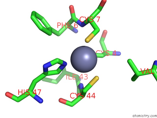
Mono view
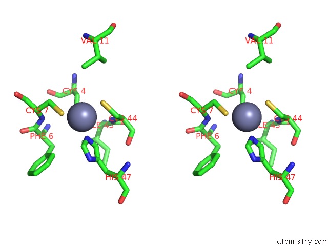
Stereo pair view
Mono view
Stereo pair view
A full contact list of Zinc with other atoms in the Zn binding
site number 1 of Crystal Structure of the Thap Domain From D. Melanogaster P-Element Transposase in Complex with Its Natural Dna Binding Site within 5.0Å range:
|
Reference:
A.Sabogal,
A.Y.Lyubimov,
J.E.Corn,
J.M.Berger,
D.C.Rio.
Thap Proteins Target Specific Dna Sites Through Bipartite Recognition of Adjacent Major and Minor Grooves. Nat.Struct.Mol.Biol. V. 17 117 2010.
ISSN: ISSN 1545-9993
PubMed: 20010837
DOI: 10.1038/NSMB.1742
Page generated: Sat Oct 26 07:45:56 2024
ISSN: ISSN 1545-9993
PubMed: 20010837
DOI: 10.1038/NSMB.1742
Last articles
Mg in 7OGNMg in 7OHN
Mg in 7OHK
Mg in 7OHG
Mg in 7OH7
Mg in 7OH6
Mg in 7OH5
Mg in 7OH4
Mg in 7OFL
Mg in 7OGF