Zinc »
PDB 3iqx-3jr3 »
3iuf »
Zinc in PDB 3iuf: Crystal Structure of the C2H2-Type Zinc Finger Domain of Human Ubi-D4
Protein crystallography data
The structure of Crystal Structure of the C2H2-Type Zinc Finger Domain of Human Ubi-D4, PDB code: 3iuf
was solved by
W.Tempel,
C.Xu,
C.Bian,
M.Adams-Cioaba,
J.Eryilmaz,
C.Bountra,
J.Weigelt,
C.H.Arrowsmith,
A.M.Edwards,
A.Bochkarev,
J.Min,
Structural Genomicsconsortium (Sgc),
with X-Ray Crystallography technique. A brief refinement statistics is given in the table below:
| Resolution Low / High (Å) | 22.72 / 1.80 |
| Space group | P 21 21 2 |
| Cell size a, b, c (Å), α, β, γ (°) | 24.714, 57.657, 22.585, 90.00, 90.00, 90.00 |
| R / Rfree (%) | 19.3 / 23.7 |
Zinc Binding Sites:
The binding sites of Zinc atom in the Crystal Structure of the C2H2-Type Zinc Finger Domain of Human Ubi-D4
(pdb code 3iuf). This binding sites where shown within
5.0 Angstroms radius around Zinc atom.
In total only one binding site of Zinc was determined in the Crystal Structure of the C2H2-Type Zinc Finger Domain of Human Ubi-D4, PDB code: 3iuf:
In total only one binding site of Zinc was determined in the Crystal Structure of the C2H2-Type Zinc Finger Domain of Human Ubi-D4, PDB code: 3iuf:
Zinc binding site 1 out of 1 in 3iuf
Go back to
Zinc binding site 1 out
of 1 in the Crystal Structure of the C2H2-Type Zinc Finger Domain of Human Ubi-D4
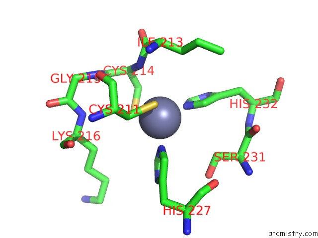
Mono view
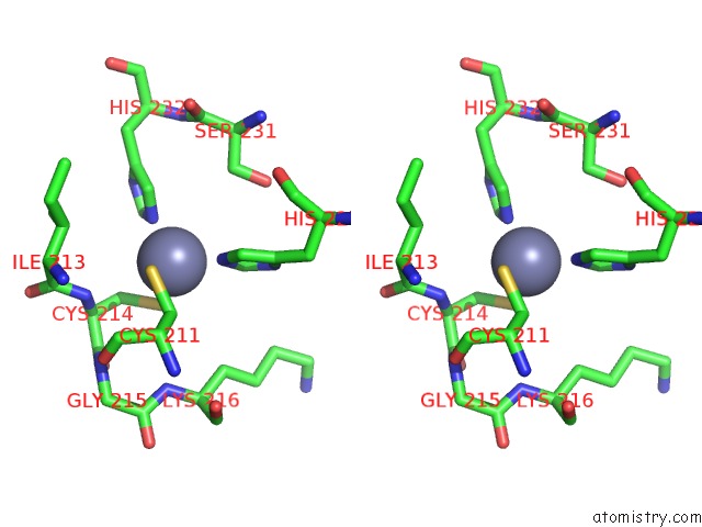
Stereo pair view
Mono view
Stereo pair view
A full contact list of Zinc with other atoms in the Zn binding
site number 1 of Crystal Structure of the C2H2-Type Zinc Finger Domain of Human Ubi-D4 within 5.0Å range:
|
Reference:
W.Zhang,
C.Xu,
C.Bian,
W.Tempel,
L.Crombet,
F.Mackenzie,
J.Min,
Z.Liu,
C.Qi.
Crystal Structure of the CYS2HIS2-Type Zinc Finger Domain of Human DPF2. Biochem.Biophys.Res.Commun. V. 413 58 2011.
ISSN: ISSN 0006-291X
PubMed: 21888896
DOI: 10.1016/J.BBRC.2011.08.043
Page generated: Sat Oct 26 07:20:59 2024
ISSN: ISSN 0006-291X
PubMed: 21888896
DOI: 10.1016/J.BBRC.2011.08.043
Last articles
Na in 9LXLNa in 9LNQ
Na in 9LS8
Na in 9LML
Na in 9LMK
Na in 9LJ6
Na in 9L6Z
Na in 9LKS
Na in 9L71
Na in 9KYO