Zinc »
PDB 3hqz-3i4n »
3i2d »
Zinc in PDB 3i2d: Crystal Structure of S. Cerevisiae Sumo E3 Ligase SIZ1
Protein crystallography data
The structure of Crystal Structure of S. Cerevisiae Sumo E3 Ligase SIZ1, PDB code: 3i2d
was solved by
C.D.Lima,
A.A.Yunus,
with X-Ray Crystallography technique. A brief refinement statistics is given in the table below:
| Resolution Low / High (Å) | 34.02 / 2.60 |
| Space group | P 21 21 21 |
| Cell size a, b, c (Å), α, β, γ (°) | 44.487, 88.980, 105.598, 90.00, 90.00, 90.00 |
| R / Rfree (%) | 21.1 / 25.9 |
Zinc Binding Sites:
The binding sites of Zinc atom in the Crystal Structure of S. Cerevisiae Sumo E3 Ligase SIZ1
(pdb code 3i2d). This binding sites where shown within
5.0 Angstroms radius around Zinc atom.
In total only one binding site of Zinc was determined in the Crystal Structure of S. Cerevisiae Sumo E3 Ligase SIZ1, PDB code: 3i2d:
In total only one binding site of Zinc was determined in the Crystal Structure of S. Cerevisiae Sumo E3 Ligase SIZ1, PDB code: 3i2d:
Zinc binding site 1 out of 1 in 3i2d
Go back to
Zinc binding site 1 out
of 1 in the Crystal Structure of S. Cerevisiae Sumo E3 Ligase SIZ1
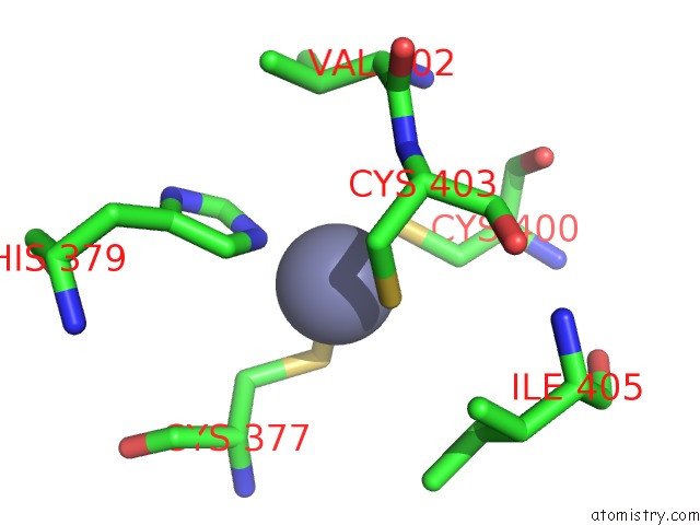
Mono view
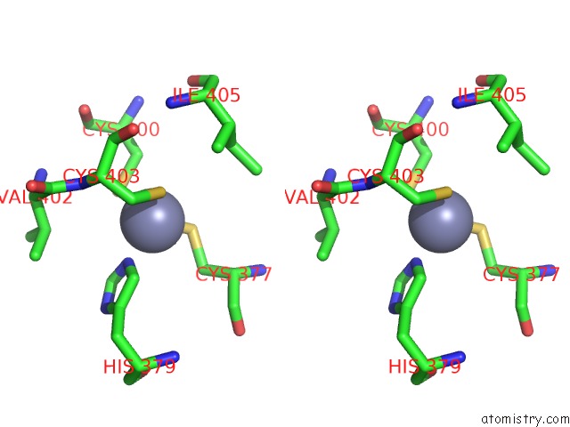
Stereo pair view
Mono view
Stereo pair view
A full contact list of Zinc with other atoms in the Zn binding
site number 1 of Crystal Structure of S. Cerevisiae Sumo E3 Ligase SIZ1 within 5.0Å range:
|
Reference:
A.A.Yunus,
C.D.Lima.
Structure of the Siz/Pias Sumo E3 Ligase SIZ1 and Determinants Required For Sumo Modification of Pcna. Mol.Cell V. 35 669 2009.
ISSN: ISSN 1097-2765
PubMed: 19748360
DOI: 10.1016/J.MOLCEL.2009.07.013
Page generated: Sat Oct 26 06:47:48 2024
ISSN: ISSN 1097-2765
PubMed: 19748360
DOI: 10.1016/J.MOLCEL.2009.07.013
Last articles
Fe in 2YXOFe in 2YRS
Fe in 2YXC
Fe in 2YNM
Fe in 2YVJ
Fe in 2YP1
Fe in 2YU2
Fe in 2YU1
Fe in 2YQB
Fe in 2YOO