Zinc »
PDB 3g8u-3gkl »
3g9y »
Zinc in PDB 3g9y: Crystal Structure of the Second Zinc Finger From ZRANB2/ZNF265 Bound to 6 Nt Ssrna Sequence Agguaa
Protein crystallography data
The structure of Crystal Structure of the Second Zinc Finger From ZRANB2/ZNF265 Bound to 6 Nt Ssrna Sequence Agguaa, PDB code: 3g9y
was solved by
F.E.Loughlin,
A.P.Mcgrath,
M.Lee,
J.M.Guss,
J.P.Mackay,
with X-Ray Crystallography technique. A brief refinement statistics is given in the table below:
| Resolution Low / High (Å) | 33.69 / 1.40 |
| Space group | P 65 2 2 |
| Cell size a, b, c (Å), α, β, γ (°) | 54.519, 54.519, 48.071, 90.00, 90.00, 120.00 |
| R / Rfree (%) | 20.1 / 23.5 |
Zinc Binding Sites:
The binding sites of Zinc atom in the Crystal Structure of the Second Zinc Finger From ZRANB2/ZNF265 Bound to 6 Nt Ssrna Sequence Agguaa
(pdb code 3g9y). This binding sites where shown within
5.0 Angstroms radius around Zinc atom.
In total only one binding site of Zinc was determined in the Crystal Structure of the Second Zinc Finger From ZRANB2/ZNF265 Bound to 6 Nt Ssrna Sequence Agguaa, PDB code: 3g9y:
In total only one binding site of Zinc was determined in the Crystal Structure of the Second Zinc Finger From ZRANB2/ZNF265 Bound to 6 Nt Ssrna Sequence Agguaa, PDB code: 3g9y:
Zinc binding site 1 out of 1 in 3g9y
Go back to
Zinc binding site 1 out
of 1 in the Crystal Structure of the Second Zinc Finger From ZRANB2/ZNF265 Bound to 6 Nt Ssrna Sequence Agguaa
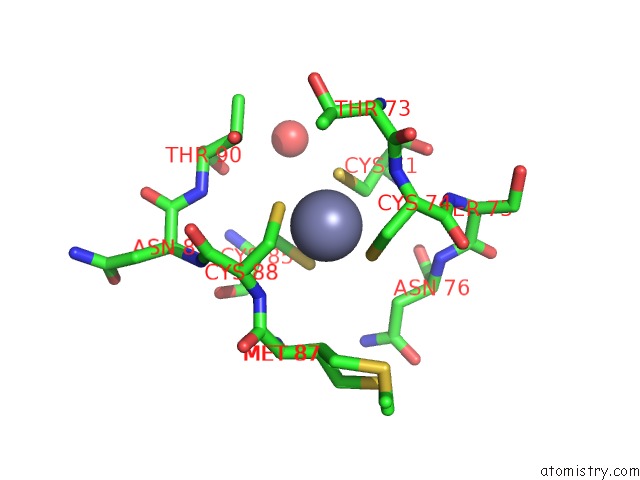
Mono view
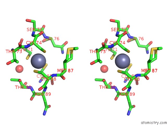
Stereo pair view
Mono view
Stereo pair view
A full contact list of Zinc with other atoms in the Zn binding
site number 1 of Crystal Structure of the Second Zinc Finger From ZRANB2/ZNF265 Bound to 6 Nt Ssrna Sequence Agguaa within 5.0Å range:
|
Reference:
F.E.Loughlin,
R.E.Mansfield,
P.M.Vaz,
A.P.Mcgrath,
S.Setiyaputra,
R.Gamsjaeger,
E.S.Chen,
B.J.Morris,
J.M.Guss,
J.P.Mackay.
The Zinc Fingers of the Sr-Like Protein ZRANB2 Are Single-Stranded Rna-Binding Domains That Recognize 5' Splice Site-Like Sequences Proc.Natl.Acad.Sci.Usa V. 106 5581 2009.
ISSN: ISSN 0027-8424
PubMed: 19304800
DOI: 10.1073/PNAS.0802466106
Page generated: Thu Oct 24 13:39:35 2024
ISSN: ISSN 0027-8424
PubMed: 19304800
DOI: 10.1073/PNAS.0802466106
Last articles
Fe in 2YXOFe in 2YRS
Fe in 2YXC
Fe in 2YNM
Fe in 2YVJ
Fe in 2YP1
Fe in 2YU2
Fe in 2YU1
Fe in 2YQB
Fe in 2YOO