Zinc »
PDB 3fum-3g8u »
3g64 »
Zinc in PDB 3g64: Crystal Structure of Putative Enoyl-Coa Hydratase From Streptomyces Coelicolor A3(2)
Protein crystallography data
The structure of Crystal Structure of Putative Enoyl-Coa Hydratase From Streptomyces Coelicolor A3(2), PDB code: 3g64
was solved by
Y.Kim,
X.Xu,
H.Cui,
A.Savchenko,
A.M.Edwards,
A.Joachimiak,
Midwest Centerfor Structural Genomics (Mcsg),
with X-Ray Crystallography technique. A brief refinement statistics is given in the table below:
| Resolution Low / High (Å) | 49.60 / 2.05 |
| Space group | C 2 2 21 |
| Cell size a, b, c (Å), α, β, γ (°) | 116.167, 191.297, 93.723, 90.00, 90.00, 90.00 |
| R / Rfree (%) | 15.4 / 19.4 |
Zinc Binding Sites:
The binding sites of Zinc atom in the Crystal Structure of Putative Enoyl-Coa Hydratase From Streptomyces Coelicolor A3(2)
(pdb code 3g64). This binding sites where shown within
5.0 Angstroms radius around Zinc atom.
In total 2 binding sites of Zinc where determined in the Crystal Structure of Putative Enoyl-Coa Hydratase From Streptomyces Coelicolor A3(2), PDB code: 3g64:
Jump to Zinc binding site number: 1; 2;
In total 2 binding sites of Zinc where determined in the Crystal Structure of Putative Enoyl-Coa Hydratase From Streptomyces Coelicolor A3(2), PDB code: 3g64:
Jump to Zinc binding site number: 1; 2;
Zinc binding site 1 out of 2 in 3g64
Go back to
Zinc binding site 1 out
of 2 in the Crystal Structure of Putative Enoyl-Coa Hydratase From Streptomyces Coelicolor A3(2)
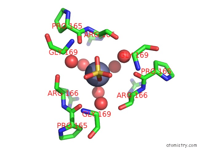
Mono view
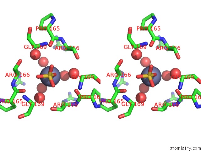
Stereo pair view
Mono view
Stereo pair view
A full contact list of Zinc with other atoms in the Zn binding
site number 1 of Crystal Structure of Putative Enoyl-Coa Hydratase From Streptomyces Coelicolor A3(2) within 5.0Å range:
|
Zinc binding site 2 out of 2 in 3g64
Go back to
Zinc binding site 2 out
of 2 in the Crystal Structure of Putative Enoyl-Coa Hydratase From Streptomyces Coelicolor A3(2)
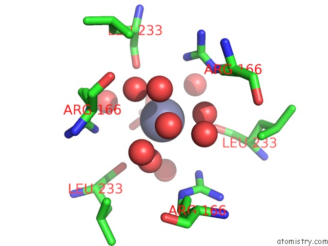
Mono view
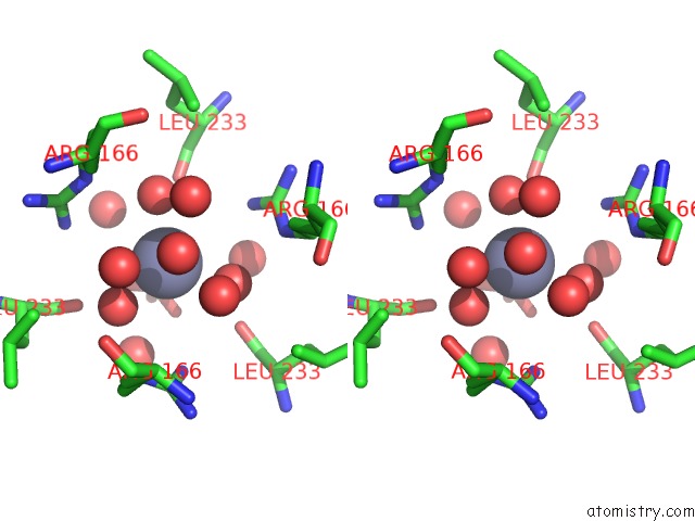
Stereo pair view
Mono view
Stereo pair view
A full contact list of Zinc with other atoms in the Zn binding
site number 2 of Crystal Structure of Putative Enoyl-Coa Hydratase From Streptomyces Coelicolor A3(2) within 5.0Å range:
|
Reference:
Y.Kim,
X.Xu,
H.Cui,
A.Savchenko,
A.M.Edwards,
A.Joachimiak.
Crystal Structure of Putative Enoyl-Coa Hydratase From Streptomyces Coelicolor A3(2) To Be Published.
Page generated: Wed Aug 20 09:26:00 2025
Last articles
Zn in 4A3EZn in 4A3C
Zn in 4A3D
Zn in 4A3B
Zn in 4A2X
Zn in 4A39
Zn in 4A38
Zn in 4A37
Zn in 4A24
Zn in 4A2C