Zinc »
PDB 3fum-3g8u »
3g4h »
Zinc in PDB 3g4h: Crystal Structure of Human Senescence Marker Protein-30 (Zinc Bound)
Protein crystallography data
The structure of Crystal Structure of Human Senescence Marker Protein-30 (Zinc Bound), PDB code: 3g4h
was solved by
S.Chakraborti,
B.J.Bahnson,
with X-Ray Crystallography technique. A brief refinement statistics is given in the table below:
| Resolution Low / High (Å) | 84.52 / 1.92 |
| Space group | P 1 21 1 |
| Cell size a, b, c (Å), α, β, γ (°) | 64.365, 52.019, 85.832, 90.00, 100.11, 90.00 |
| R / Rfree (%) | 22.4 / 25.3 |
Zinc Binding Sites:
The binding sites of Zinc atom in the Crystal Structure of Human Senescence Marker Protein-30 (Zinc Bound)
(pdb code 3g4h). This binding sites where shown within
5.0 Angstroms radius around Zinc atom.
In total 2 binding sites of Zinc where determined in the Crystal Structure of Human Senescence Marker Protein-30 (Zinc Bound), PDB code: 3g4h:
Jump to Zinc binding site number: 1; 2;
In total 2 binding sites of Zinc where determined in the Crystal Structure of Human Senescence Marker Protein-30 (Zinc Bound), PDB code: 3g4h:
Jump to Zinc binding site number: 1; 2;
Zinc binding site 1 out of 2 in 3g4h
Go back to
Zinc binding site 1 out
of 2 in the Crystal Structure of Human Senescence Marker Protein-30 (Zinc Bound)
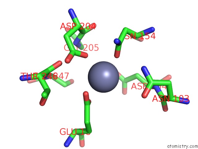
Mono view
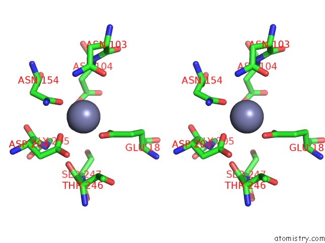
Stereo pair view
Mono view
Stereo pair view
A full contact list of Zinc with other atoms in the Zn binding
site number 1 of Crystal Structure of Human Senescence Marker Protein-30 (Zinc Bound) within 5.0Å range:
|
Zinc binding site 2 out of 2 in 3g4h
Go back to
Zinc binding site 2 out
of 2 in the Crystal Structure of Human Senescence Marker Protein-30 (Zinc Bound)
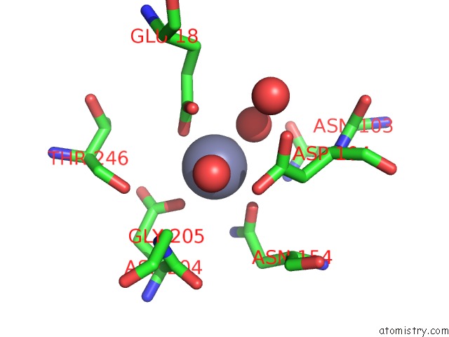
Mono view
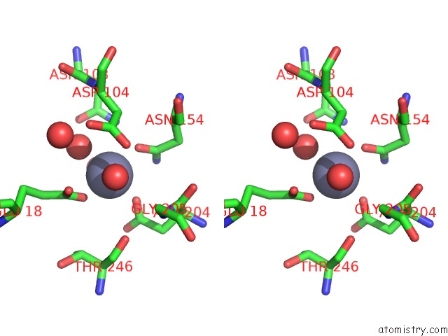
Stereo pair view
Mono view
Stereo pair view
A full contact list of Zinc with other atoms in the Zn binding
site number 2 of Crystal Structure of Human Senescence Marker Protein-30 (Zinc Bound) within 5.0Å range:
|
Reference:
S.Chakraborti,
B.J.Bahnson.
Crystal Structure of Human Senescence Marker Protein 30: Insights Linking Structural, Enzymatic, and Physiological Functions . Biochemistry V. 49 3436 2010.
ISSN: ISSN 0006-2960
PubMed: 20329768
DOI: 10.1021/BI9022297
Page generated: Wed Aug 20 09:24:33 2025
ISSN: ISSN 0006-2960
PubMed: 20329768
DOI: 10.1021/BI9022297
Last articles
Zn in 4A47Zn in 4A46
Zn in 4A3W
Zn in 4A3N
Zn in 4A3L
Zn in 4A3J
Zn in 4A3M
Zn in 4A3K
Zn in 4A3G
Zn in 4A3I