Zinc »
PDB 3ful-3g8r »
3g27 »
Zinc in PDB 3g27: Structure of A Putative Bacteriophage Protein From Escherichia Coli Str. K-12 Substr. MG1655
Protein crystallography data
The structure of Structure of A Putative Bacteriophage Protein From Escherichia Coli Str. K-12 Substr. MG1655, PDB code: 3g27
was solved by
M.E.Cuff,
E.Evdokimova,
M.Kudritska,
A.Edwards,
A.Savchenko,
A.Joachimiak,
Midwest Center For Structural Genomics (Mcsg),
with X-Ray Crystallography technique. A brief refinement statistics is given in the table below:
| Resolution Low / High (Å) | 30.88 / 2.10 |
| Space group | P 31 2 1 |
| Cell size a, b, c (Å), α, β, γ (°) | 71.311, 71.311, 32.333, 90.00, 90.00, 120.00 |
| R / Rfree (%) | 18.6 / 24.9 |
Other elements in 3g27:
The structure of Structure of A Putative Bacteriophage Protein From Escherichia Coli Str. K-12 Substr. MG1655 also contains other interesting chemical elements:
| Calcium | (Ca) | 1 atom |
Zinc Binding Sites:
The binding sites of Zinc atom in the Structure of A Putative Bacteriophage Protein From Escherichia Coli Str. K-12 Substr. MG1655
(pdb code 3g27). This binding sites where shown within
5.0 Angstroms radius around Zinc atom.
In total only one binding site of Zinc was determined in the Structure of A Putative Bacteriophage Protein From Escherichia Coli Str. K-12 Substr. MG1655, PDB code: 3g27:
In total only one binding site of Zinc was determined in the Structure of A Putative Bacteriophage Protein From Escherichia Coli Str. K-12 Substr. MG1655, PDB code: 3g27:
Zinc binding site 1 out of 1 in 3g27
Go back to
Zinc binding site 1 out
of 1 in the Structure of A Putative Bacteriophage Protein From Escherichia Coli Str. K-12 Substr. MG1655
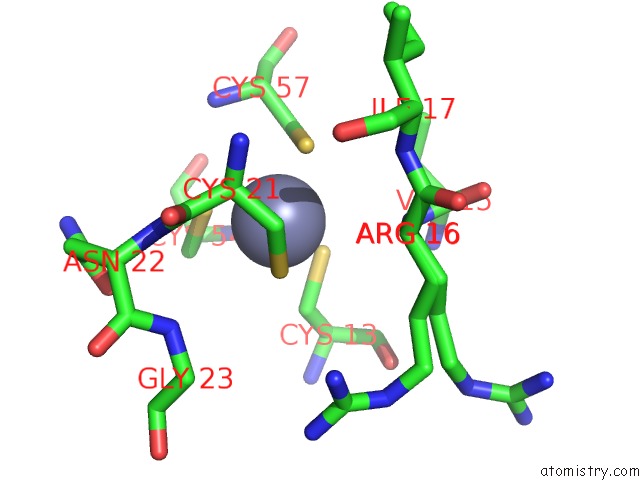
Mono view
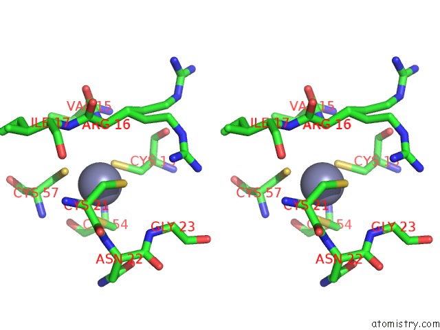
Stereo pair view
Mono view
Stereo pair view
A full contact list of Zinc with other atoms in the Zn binding
site number 1 of Structure of A Putative Bacteriophage Protein From Escherichia Coli Str. K-12 Substr. MG1655 within 5.0Å range:
|
Reference:
M.E.Cuff,
E.Evdokimova,
M.Kudritska,
A.Edwards,
A.Savchenko,
A.Joachimiak.
Structure of A Putative Bacteriophage Protein From Escherichia Coli Str. K-12 Substr. MG1655 To Be Published.
Page generated: Thu Oct 24 13:28:58 2024
Last articles
Na in 8GT0Na in 8H2B
Na in 8GZ1
Na in 8H1G
Na in 8H1B
Na in 8GYR
Na in 8GVM
Na in 8GR6
Na in 8GQ0
Na in 8GSG