Zinc »
PDB 3flf-3fuk »
3fp5 »
Zinc in PDB 3fp5: Crystal Structure of Acbp From Moniliophthora Perniciosa
Protein crystallography data
The structure of Crystal Structure of Acbp From Moniliophthora Perniciosa, PDB code: 3fp5
was solved by
P.S.Monzani,
H.M.Pereira,
F.A.Melo,
F.V.Meirelles,
G.Oliva,
J.C.M.Cascardo,
with X-Ray Crystallography technique. A brief refinement statistics is given in the table below:
| Resolution Low / High (Å) | 18.68 / 1.61 |
| Space group | P 21 21 21 |
| Cell size a, b, c (Å), α, β, γ (°) | 32.500, 41.024, 82.463, 90.00, 90.00, 90.00 |
| R / Rfree (%) | 18.6 / 21.2 |
Zinc Binding Sites:
The binding sites of Zinc atom in the Crystal Structure of Acbp From Moniliophthora Perniciosa
(pdb code 3fp5). This binding sites where shown within
5.0 Angstroms radius around Zinc atom.
In total only one binding site of Zinc was determined in the Crystal Structure of Acbp From Moniliophthora Perniciosa, PDB code: 3fp5:
In total only one binding site of Zinc was determined in the Crystal Structure of Acbp From Moniliophthora Perniciosa, PDB code: 3fp5:
Zinc binding site 1 out of 1 in 3fp5
Go back to
Zinc binding site 1 out
of 1 in the Crystal Structure of Acbp From Moniliophthora Perniciosa
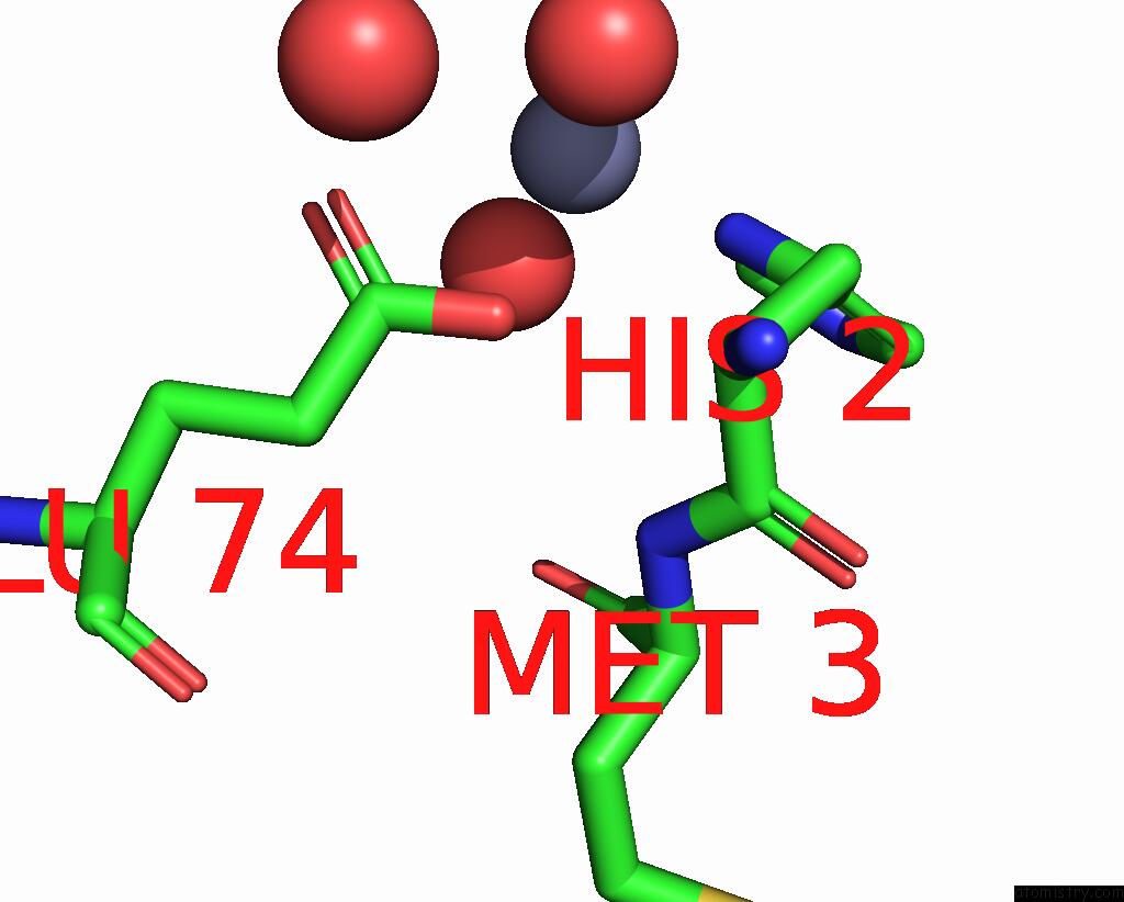
Mono view
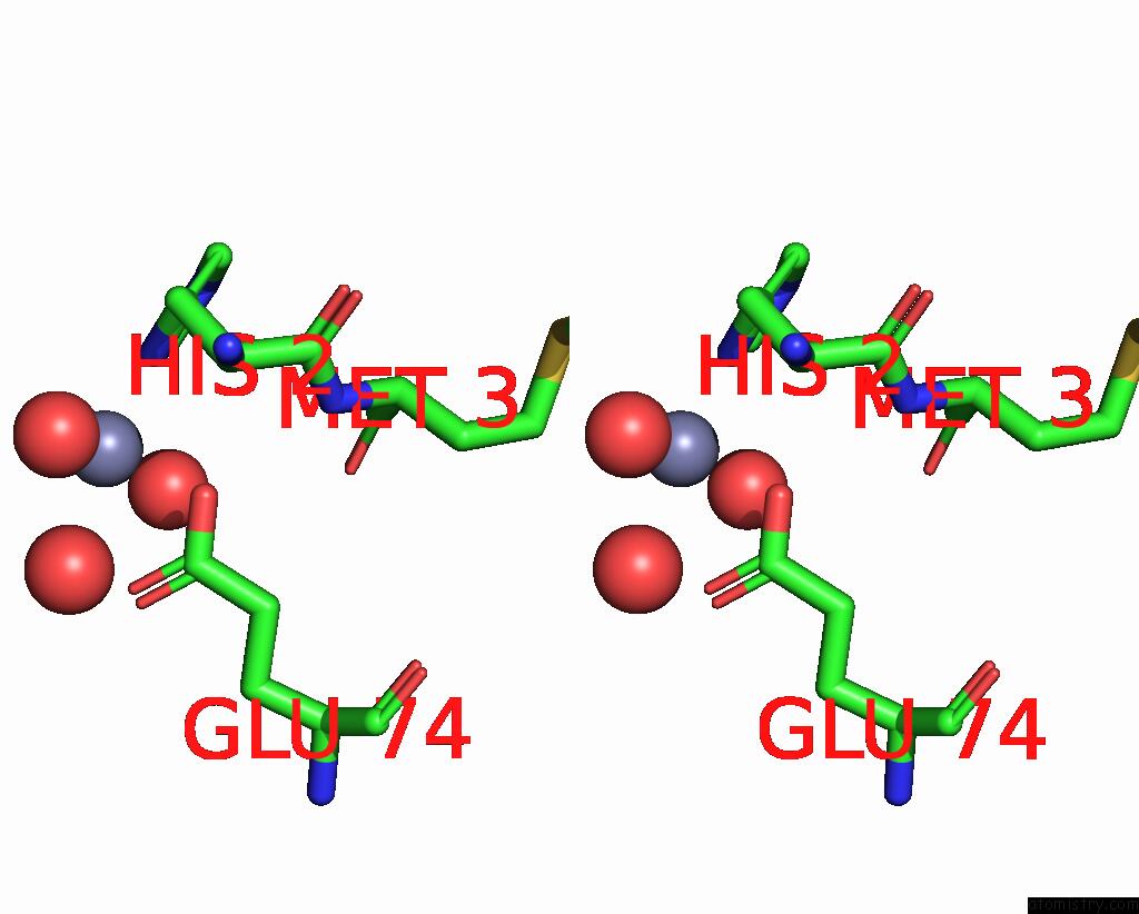
Stereo pair view
Mono view
Stereo pair view
A full contact list of Zinc with other atoms in the Zn binding
site number 1 of Crystal Structure of Acbp From Moniliophthora Perniciosa within 5.0Å range:
|
Reference:
P.S.Monzani,
H.M.Pereira,
F.A.Melo,
F.V.Meirelles,
G.Oliva,
J.C.Cascardo.
A New Topology of Acbp From Moniliophthora Perniciosa. Biochim.Biophys.Acta V.1804 115 2010.
ISSN: ISSN 0006-3002
PubMed: 19782157
DOI: 10.1016/J.BBAPAP.2009.09.020
Page generated: Thu Oct 24 13:20:58 2024
ISSN: ISSN 0006-3002
PubMed: 19782157
DOI: 10.1016/J.BBAPAP.2009.09.020
Last articles
Mg in 3T2CMg in 3T2B
Mg in 3T1R
Mg in 3T1O
Mg in 3T0D
Mg in 3T1Q
Mg in 3T12
Mg in 3T1K
Mg in 3T10
Mg in 3T0Z