Zinc »
PDB 3ekz-3eyv »
3ewf »
Zinc in PDB 3ewf: Crystal Structure Analysis of Human HDAC8 H143A Variant Complexed with Substrate.
Enzymatic activity of Crystal Structure Analysis of Human HDAC8 H143A Variant Complexed with Substrate.
All present enzymatic activity of Crystal Structure Analysis of Human HDAC8 H143A Variant Complexed with Substrate.:
3.5.1.98;
3.5.1.98;
Protein crystallography data
The structure of Crystal Structure Analysis of Human HDAC8 H143A Variant Complexed with Substrate., PDB code: 3ewf
was solved by
D.P.Dowling,
S.L.Gantt,
S.G.Gattis,
C.A.Fierke,
D.W.Christianon,
with X-Ray Crystallography technique. A brief refinement statistics is given in the table below:
| Resolution Low / High (Å) | 49.15 / 2.50 |
| Space group | P 21 21 21 |
| Cell size a, b, c (Å), α, β, γ (°) | 82.903, 91.844, 196.577, 90.00, 90.00, 90.00 |
| R / Rfree (%) | 19.8 / 22.9 |
Other elements in 3ewf:
The structure of Crystal Structure Analysis of Human HDAC8 H143A Variant Complexed with Substrate. also contains other interesting chemical elements:
| Potassium | (K) | 8 atoms |
Zinc Binding Sites:
The binding sites of Zinc atom in the Crystal Structure Analysis of Human HDAC8 H143A Variant Complexed with Substrate.
(pdb code 3ewf). This binding sites where shown within
5.0 Angstroms radius around Zinc atom.
In total 6 binding sites of Zinc where determined in the Crystal Structure Analysis of Human HDAC8 H143A Variant Complexed with Substrate., PDB code: 3ewf:
Jump to Zinc binding site number: 1; 2; 3; 4; 5; 6;
In total 6 binding sites of Zinc where determined in the Crystal Structure Analysis of Human HDAC8 H143A Variant Complexed with Substrate., PDB code: 3ewf:
Jump to Zinc binding site number: 1; 2; 3; 4; 5; 6;
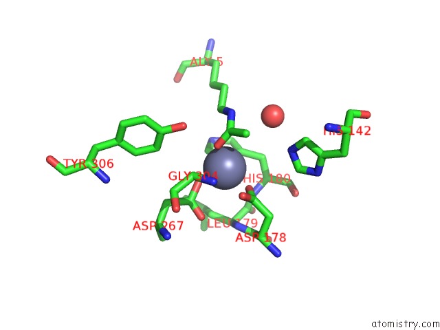
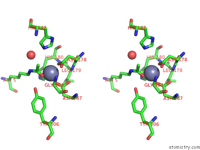
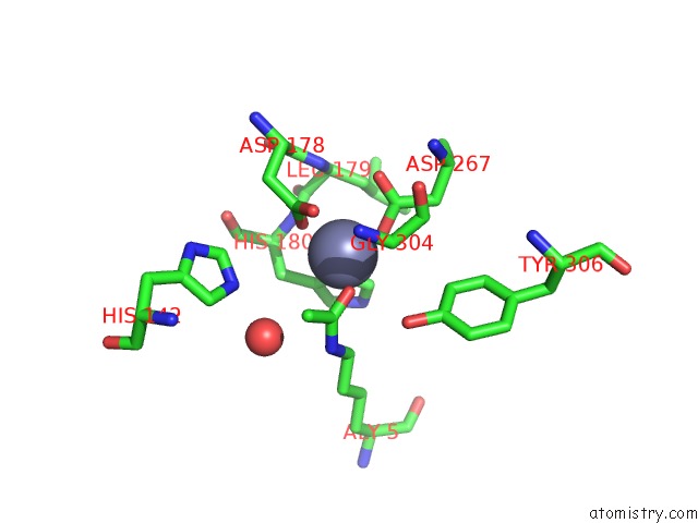
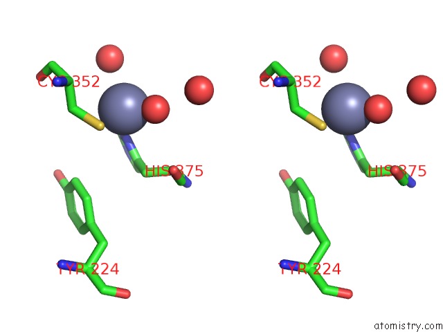
Zinc binding site 1 out of 6 in 3ewf
Go back to
Zinc binding site 1 out
of 6 in the Crystal Structure Analysis of Human HDAC8 H143A Variant Complexed with Substrate.
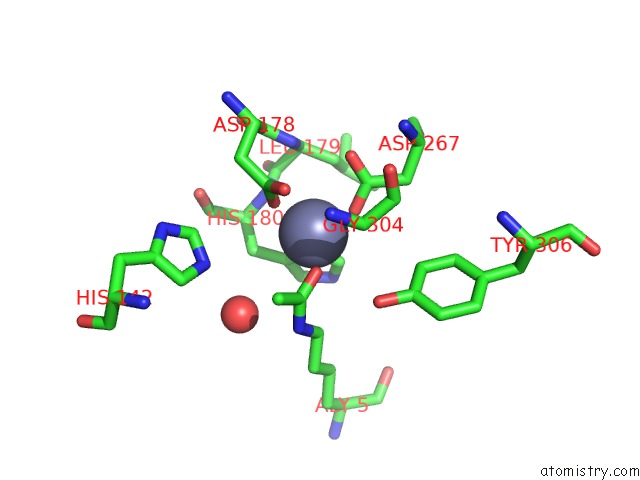
Mono view
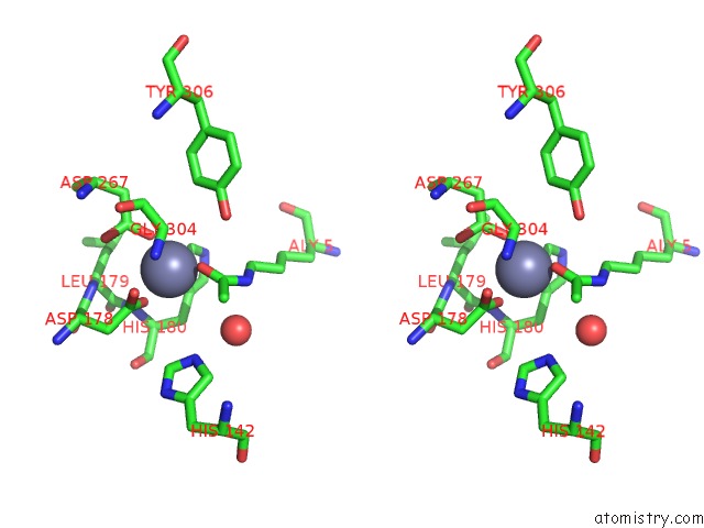
Stereo pair view
Mono view
Stereo pair view
A full contact list of Zinc with other atoms in the Zn binding
site number 1 of Crystal Structure Analysis of Human HDAC8 H143A Variant Complexed with Substrate. within 5.0Å range:
|
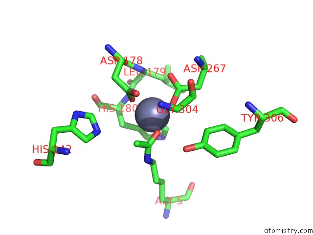
Zinc binding site 2 out of 6 in 3ewf
Go back to
Zinc binding site 2 out
of 6 in the Crystal Structure Analysis of Human HDAC8 H143A Variant Complexed with Substrate.
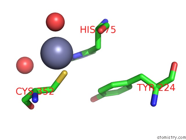
Mono view
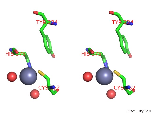
Stereo pair view
Mono view
Stereo pair view
A full contact list of Zinc with other atoms in the Zn binding
site number 2 of Crystal Structure Analysis of Human HDAC8 H143A Variant Complexed with Substrate. within 5.0Å range:
|
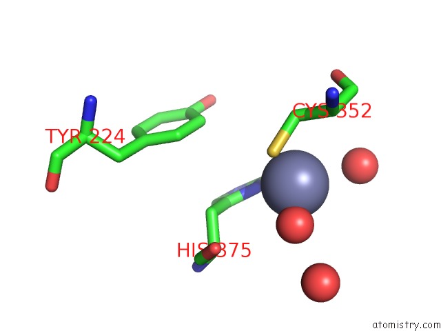
Zinc binding site 3 out of 6 in 3ewf
Go back to
Zinc binding site 3 out
of 6 in the Crystal Structure Analysis of Human HDAC8 H143A Variant Complexed with Substrate.
Mono view
Stereo pair view
Mono view
Stereo pair view
A full contact list of Zinc with other atoms in the Zn binding
site number 3 of Crystal Structure Analysis of Human HDAC8 H143A Variant Complexed with Substrate. within 5.0Å range:
|
Zinc binding site 4 out of 6 in 3ewf
Go back to
Zinc binding site 4 out
of 6 in the Crystal Structure Analysis of Human HDAC8 H143A Variant Complexed with Substrate.
Mono view

Stereo pair view
Mono view
Stereo pair view
A full contact list of Zinc with other atoms in the Zn binding
site number 4 of Crystal Structure Analysis of Human HDAC8 H143A Variant Complexed with Substrate. within 5.0Å range:
|
Zinc binding site 5 out of 6 in 3ewf
Go back to
Zinc binding site 5 out
of 6 in the Crystal Structure Analysis of Human HDAC8 H143A Variant Complexed with Substrate.
Mono view
Stereo pair view
Mono view
Stereo pair view
A full contact list of Zinc with other atoms in the Zn binding
site number 5 of Crystal Structure Analysis of Human HDAC8 H143A Variant Complexed with Substrate. within 5.0Å range:
|
Zinc binding site 6 out of 6 in 3ewf
Go back to
Zinc binding site 6 out
of 6 in the Crystal Structure Analysis of Human HDAC8 H143A Variant Complexed with Substrate.
Mono view

Stereo pair view
Mono view
Stereo pair view
A full contact list of Zinc with other atoms in the Zn binding
site number 6 of Crystal Structure Analysis of Human HDAC8 H143A Variant Complexed with Substrate. within 5.0Å range:
|
Reference:
D.P.Dowling,
S.L.Gantt,
S.G.Gattis,
C.A.Fierke,
D.W.Christianson.
Structural Studies of Human Histone Deacetylase 8 and Its Site-Specific Variants Complexed with Substrate and Inhibitors. Biochemistry V. 47 13554 2008.
ISSN: ISSN 0006-2960
PubMed: 19053282
DOI: 10.1021/BI801610C
Page generated: Thu Oct 24 12:56:49 2024
ISSN: ISSN 0006-2960
PubMed: 19053282
DOI: 10.1021/BI801610C
Last articles
Mg in 9IUYMg in 9IXM
Mg in 9IXF
Mg in 9IXG
Mg in 9IWG
Mg in 9IXE
Mg in 9IXD
Mg in 9IXC
Mg in 9IWT
Mg in 9IWF