Zinc »
PDB 3e2w-3ebe »
3eb5 »
Zinc in PDB 3eb5: Structure of the CIAP2 Ring Domain
Protein crystallography data
The structure of Structure of the CIAP2 Ring Domain, PDB code: 3eb5
was solved by
P.D.Mace,
K.Linke,
C.A.Smith,
C.L.Day,
with X-Ray Crystallography technique. A brief refinement statistics is given in the table below:
| Resolution Low / High (Å) | 28.69 / 2.00 |
| Space group | C 2 2 2 |
| Cell size a, b, c (Å), α, β, γ (°) | 34.869, 66.474, 77.465, 90.00, 90.00, 90.00 |
| R / Rfree (%) | 21.3 / 27.5 |
Other elements in 3eb5:
The structure of Structure of the CIAP2 Ring Domain also contains other interesting chemical elements:
| Sodium | (Na) | 1 atom |
Zinc Binding Sites:
The binding sites of Zinc atom in the Structure of the CIAP2 Ring Domain
(pdb code 3eb5). This binding sites where shown within
5.0 Angstroms radius around Zinc atom.
In total 2 binding sites of Zinc where determined in the Structure of the CIAP2 Ring Domain, PDB code: 3eb5:
Jump to Zinc binding site number: 1; 2;
In total 2 binding sites of Zinc where determined in the Structure of the CIAP2 Ring Domain, PDB code: 3eb5:
Jump to Zinc binding site number: 1; 2;
Zinc binding site 1 out of 2 in 3eb5
Go back to
Zinc binding site 1 out
of 2 in the Structure of the CIAP2 Ring Domain
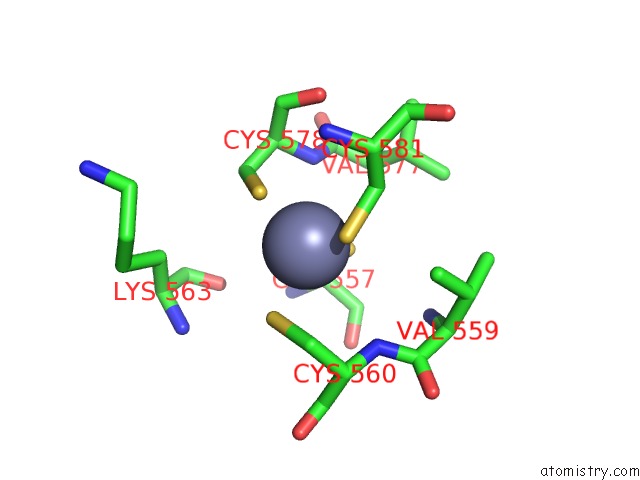
Mono view
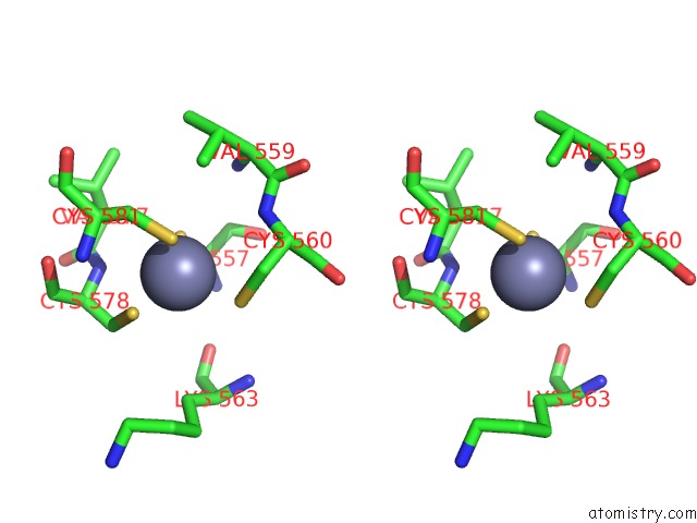
Stereo pair view
Mono view
Stereo pair view
A full contact list of Zinc with other atoms in the Zn binding
site number 1 of Structure of the CIAP2 Ring Domain within 5.0Å range:
|
Zinc binding site 2 out of 2 in 3eb5
Go back to
Zinc binding site 2 out
of 2 in the Structure of the CIAP2 Ring Domain
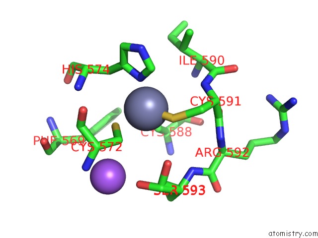
Mono view
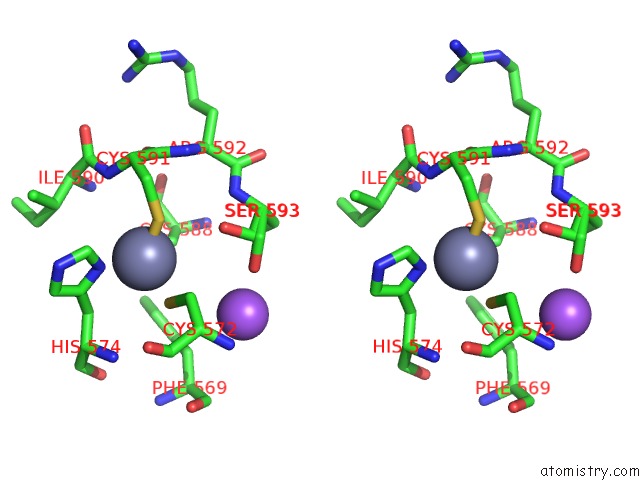
Stereo pair view
Mono view
Stereo pair view
A full contact list of Zinc with other atoms in the Zn binding
site number 2 of Structure of the CIAP2 Ring Domain within 5.0Å range:
|
Reference:
P.D.Mace,
K.Linke,
R.Feltham,
F.R.Schumacher,
C.A.Smith,
D.L.Vaux,
J.Silke,
C.L.Day.
Structures of the CIAP2 Ring Domain Reveal Conformational Changes Associated with Ubiquitin-Conjugating Enzyme (E2) Recruitment. J.Biol.Chem. V. 283 31633 2008.
ISSN: ISSN 0021-9258
PubMed: 18784070
DOI: 10.1074/JBC.M804753200
Page generated: Wed Aug 20 08:50:24 2025
ISSN: ISSN 0021-9258
PubMed: 18784070
DOI: 10.1074/JBC.M804753200
Last articles
Mn in 9LJUMn in 9LJW
Mn in 9LJS
Mn in 9LJR
Mn in 9LJT
Mn in 9LJV
Mg in 9UA2
Mg in 9R96
Mg in 9VM1
Mg in 9P01