Zinc »
PDB 3dgn-3dsw »
3dsu »
Zinc in PDB 3dsu: Crystal Structure of Rabggtase(Delta Lrr; Delta Ig)in Complex with Farnesyl Pyrophosphate
Enzymatic activity of Crystal Structure of Rabggtase(Delta Lrr; Delta Ig)in Complex with Farnesyl Pyrophosphate
All present enzymatic activity of Crystal Structure of Rabggtase(Delta Lrr; Delta Ig)in Complex with Farnesyl Pyrophosphate:
2.5.1.60;
2.5.1.60;
Protein crystallography data
The structure of Crystal Structure of Rabggtase(Delta Lrr; Delta Ig)in Complex with Farnesyl Pyrophosphate, PDB code: 3dsu
was solved by
Z.Guo,
S.Yu,
R.S.Goody,
K.Alexandrov,
W.Blankenfeldt,
with X-Ray Crystallography technique. A brief refinement statistics is given in the table below:
| Resolution Low / High (Å) | 29.19 / 1.90 |
| Space group | P 21 21 21 |
| Cell size a, b, c (Å), α, β, γ (°) | 66.875, 90.877, 114.240, 90.00, 90.00, 90.00 |
| R / Rfree (%) | 15.9 / 21.5 |
Other elements in 3dsu:
The structure of Crystal Structure of Rabggtase(Delta Lrr; Delta Ig)in Complex with Farnesyl Pyrophosphate also contains other interesting chemical elements:
| Calcium | (Ca) | 1 atom |
Zinc Binding Sites:
The binding sites of Zinc atom in the Crystal Structure of Rabggtase(Delta Lrr; Delta Ig)in Complex with Farnesyl Pyrophosphate
(pdb code 3dsu). This binding sites where shown within
5.0 Angstroms radius around Zinc atom.
In total only one binding site of Zinc was determined in the Crystal Structure of Rabggtase(Delta Lrr; Delta Ig)in Complex with Farnesyl Pyrophosphate, PDB code: 3dsu:
In total only one binding site of Zinc was determined in the Crystal Structure of Rabggtase(Delta Lrr; Delta Ig)in Complex with Farnesyl Pyrophosphate, PDB code: 3dsu:
Zinc binding site 1 out of 1 in 3dsu
Go back to
Zinc binding site 1 out
of 1 in the Crystal Structure of Rabggtase(Delta Lrr; Delta Ig)in Complex with Farnesyl Pyrophosphate

Mono view
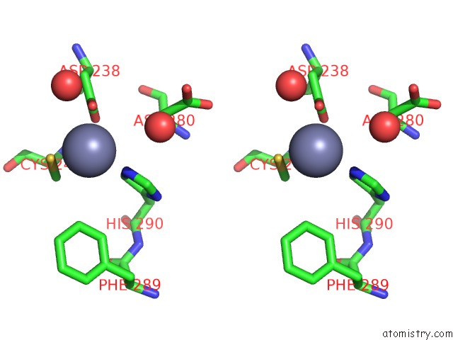
Stereo pair view
Mono view
Stereo pair view
A full contact list of Zinc with other atoms in the Zn binding
site number 1 of Crystal Structure of Rabggtase(Delta Lrr; Delta Ig)in Complex with Farnesyl Pyrophosphate within 5.0Å range:
|
Reference:
Z.Guo,
Y.-W.Wu,
D.Das,
C.Delon,
J.Cramer,
S.Yu,
S.Thuns,
N.Lupilova,
H.Waldmann,
L.Brunsveld,
R.S.Goody,
K.Alexandrov,
W.Blankenfeldt.
Structures of Rabggtase-Substrate/Product Complexes Provide Insights Into the Evolution of Protein Prenylation Embo J. V. 27 2444 2008.
ISSN: ISSN 0261-4189
PubMed: 18756270
DOI: 10.1038/EMBOJ.2008.164
Page generated: Thu Oct 24 12:19:51 2024
ISSN: ISSN 0261-4189
PubMed: 18756270
DOI: 10.1038/EMBOJ.2008.164
Last articles
Mg in 4RRIMg in 4RRF
Mg in 4RRH
Mg in 4RRD
Mg in 4RRA
Mg in 4RR9
Mg in 4RR8
Mg in 4RR7
Mg in 4RQ5
Mg in 4RQI