Zinc »
PDB 3czt-3d7s »
3d4b »
Zinc in PDB 3d4b: Crystal Structure of SIR2TM in Complex with Acetyl P53 Peptide and Dadme-Nad+
Protein crystallography data
The structure of Crystal Structure of SIR2TM in Complex with Acetyl P53 Peptide and Dadme-Nad+, PDB code: 3d4b
was solved by
W.F.Hawse,
K.G.Hoff,
D.Fatkins,
A.Daines,
O.V.Zubkova,
V.L.Schramm,
W.Zheng,
C.Wolberger,
with X-Ray Crystallography technique. A brief refinement statistics is given in the table below:
| Resolution Low / High (Å) | 50.00 / 1.90 |
| Space group | P 21 21 21 |
| Cell size a, b, c (Å), α, β, γ (°) | 45.790, 59.735, 106.114, 90.00, 90.00, 90.00 |
| R / Rfree (%) | 19.5 / 22.9 |
Zinc Binding Sites:
The binding sites of Zinc atom in the Crystal Structure of SIR2TM in Complex with Acetyl P53 Peptide and Dadme-Nad+
(pdb code 3d4b). This binding sites where shown within
5.0 Angstroms radius around Zinc atom.
In total only one binding site of Zinc was determined in the Crystal Structure of SIR2TM in Complex with Acetyl P53 Peptide and Dadme-Nad+, PDB code: 3d4b:
In total only one binding site of Zinc was determined in the Crystal Structure of SIR2TM in Complex with Acetyl P53 Peptide and Dadme-Nad+, PDB code: 3d4b:
Zinc binding site 1 out of 1 in 3d4b
Go back to
Zinc binding site 1 out
of 1 in the Crystal Structure of SIR2TM in Complex with Acetyl P53 Peptide and Dadme-Nad+
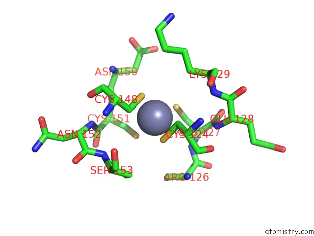
Mono view
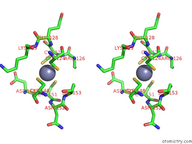
Stereo pair view
Mono view
Stereo pair view
A full contact list of Zinc with other atoms in the Zn binding
site number 1 of Crystal Structure of SIR2TM in Complex with Acetyl P53 Peptide and Dadme-Nad+ within 5.0Å range:
|
Reference:
W.F.Hawse,
K.G.Hoff,
D.G.Fatkins,
A.Daines,
O.V.Zubkova,
V.L.Schramm,
W.Zheng,
C.Wolberger.
Structural Insights Into Intermediate Steps in the SIR2 Deacetylation Reaction. Structure V. 16 1368 2008.
ISSN: ISSN 0969-2126
PubMed: 18786399
DOI: 10.1016/J.STR.2008.05.015
Page generated: Thu Oct 24 12:02:36 2024
ISSN: ISSN 0969-2126
PubMed: 18786399
DOI: 10.1016/J.STR.2008.05.015
Last articles
Fe in 8DW0Fe in 8DW7
Fe in 8DW4
Fe in 8DVY
Fe in 8DVP
Fe in 8DPN
Fe in 8DQV
Fe in 8DVX
Fe in 8DSG
Fe in 8DNP