Zinc »
PDB 3cjp-3czs »
3coq »
Zinc in PDB 3coq: Structural Basis For Dimerization in Dna Recognition By GAL4
Protein crystallography data
The structure of Structural Basis For Dimerization in Dna Recognition By GAL4, PDB code: 3coq
was solved by
M.Hong,
M.X.Fitzgerald,
S.Harper,
C.Luo,
D.W.Speicher,
with X-Ray Crystallography technique. A brief refinement statistics is given in the table below:
| Resolution Low / High (Å) | 28.94 / 2.40 |
| Space group | C 1 2 1 |
| Cell size a, b, c (Å), α, β, γ (°) | 126.495, 40.829, 90.418, 90.00, 95.88, 90.00 |
| R / Rfree (%) | 20.7 / 26.9 |
Zinc Binding Sites:
The binding sites of Zinc atom in the Structural Basis For Dimerization in Dna Recognition By GAL4
(pdb code 3coq). This binding sites where shown within
5.0 Angstroms radius around Zinc atom.
In total 4 binding sites of Zinc where determined in the Structural Basis For Dimerization in Dna Recognition By GAL4, PDB code: 3coq:
Jump to Zinc binding site number: 1; 2; 3; 4;
In total 4 binding sites of Zinc where determined in the Structural Basis For Dimerization in Dna Recognition By GAL4, PDB code: 3coq:
Jump to Zinc binding site number: 1; 2; 3; 4;
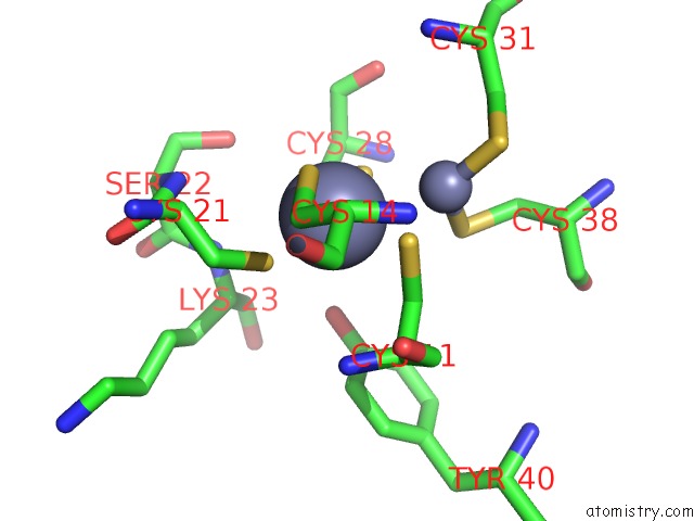
Zinc binding site 1 out of 4 in 3coq
Go back to
Zinc binding site 1 out
of 4 in the Structural Basis For Dimerization in Dna Recognition By GAL4
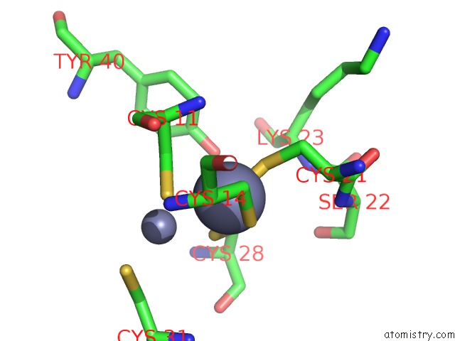
Mono view
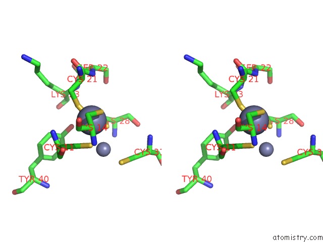
Stereo pair view
Mono view
Stereo pair view
A full contact list of Zinc with other atoms in the Zn binding
site number 1 of Structural Basis For Dimerization in Dna Recognition By GAL4 within 5.0Å range:
|
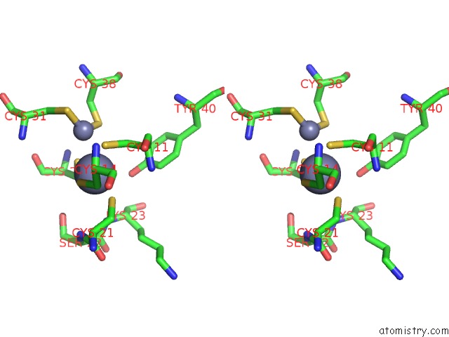
Zinc binding site 2 out of 4 in 3coq
Go back to
Zinc binding site 2 out
of 4 in the Structural Basis For Dimerization in Dna Recognition By GAL4
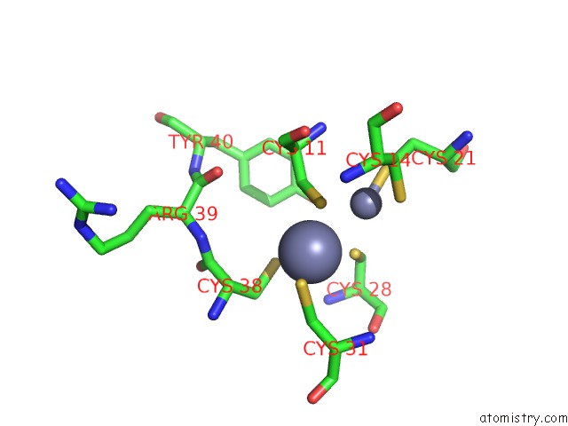
Mono view
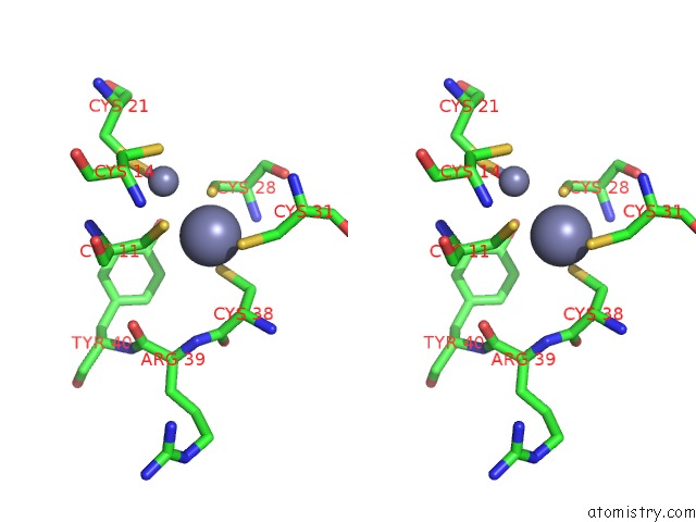
Stereo pair view
Mono view
Stereo pair view
A full contact list of Zinc with other atoms in the Zn binding
site number 2 of Structural Basis For Dimerization in Dna Recognition By GAL4 within 5.0Å range:
|
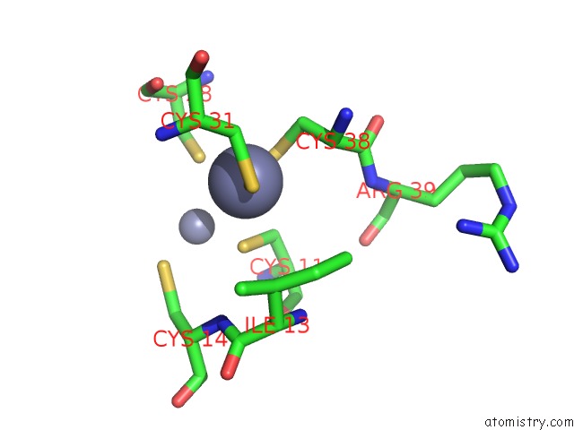
Zinc binding site 3 out of 4 in 3coq
Go back to
Zinc binding site 3 out
of 4 in the Structural Basis For Dimerization in Dna Recognition By GAL4
Mono view
Stereo pair view
Mono view
Stereo pair view
A full contact list of Zinc with other atoms in the Zn binding
site number 3 of Structural Basis For Dimerization in Dna Recognition By GAL4 within 5.0Å range:
|
Zinc binding site 4 out of 4 in 3coq
Go back to
Zinc binding site 4 out
of 4 in the Structural Basis For Dimerization in Dna Recognition By GAL4
Mono view
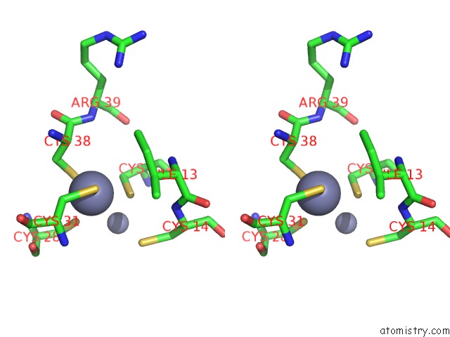
Stereo pair view
Mono view
Stereo pair view
A full contact list of Zinc with other atoms in the Zn binding
site number 4 of Structural Basis For Dimerization in Dna Recognition By GAL4 within 5.0Å range:
|
Reference:
M.Hong,
M.X.Fitzgerald,
S.Harper,
C.Luo,
D.W.Speicher,
R.Marmorstein.
Structural Basis For Dimerization in Dna Recognition By GAL4. Structure V. 16 1019 2008.
ISSN: ISSN 0969-2126
PubMed: 18611375
DOI: 10.1016/J.STR.2008.03.015
Page generated: Thu Oct 24 11:53:34 2024
ISSN: ISSN 0969-2126
PubMed: 18611375
DOI: 10.1016/J.STR.2008.03.015
Last articles
K in 7BVYK in 7BVU
K in 7BT2
K in 7BMZ
K in 7BMU
K in 7BH2
K in 7BIL
K in 7BI1
K in 7BH1
K in 7B2H