Zinc »
PDB 3b7r-3bo5 »
3bi1 »
Zinc in PDB 3bi1: X-Ray Structure of Human Glutamate Carboxypeptidase II (Gcpii) in Complex with A Transition State Analog of Methotrexate-Glu
Enzymatic activity of X-Ray Structure of Human Glutamate Carboxypeptidase II (Gcpii) in Complex with A Transition State Analog of Methotrexate-Glu
All present enzymatic activity of X-Ray Structure of Human Glutamate Carboxypeptidase II (Gcpii) in Complex with A Transition State Analog of Methotrexate-Glu:
3.4.17.21;
3.4.17.21;
Protein crystallography data
The structure of X-Ray Structure of Human Glutamate Carboxypeptidase II (Gcpii) in Complex with A Transition State Analog of Methotrexate-Glu, PDB code: 3bi1
was solved by
C.Barinka,
J.Lubkowski,
with X-Ray Crystallography technique. A brief refinement statistics is given in the table below:
| Resolution Low / High (Å) | 15.00 / 1.50 |
| Space group | I 2 2 2 |
| Cell size a, b, c (Å), α, β, γ (°) | 102.116, 130.462, 159.456, 90.00, 90.00, 90.00 |
| R / Rfree (%) | 14.8 / 16.9 |
Other elements in 3bi1:
The structure of X-Ray Structure of Human Glutamate Carboxypeptidase II (Gcpii) in Complex with A Transition State Analog of Methotrexate-Glu also contains other interesting chemical elements:
| Chlorine | (Cl) | 1 atom |
| Calcium | (Ca) | 1 atom |
Zinc Binding Sites:
The binding sites of Zinc atom in the X-Ray Structure of Human Glutamate Carboxypeptidase II (Gcpii) in Complex with A Transition State Analog of Methotrexate-Glu
(pdb code 3bi1). This binding sites where shown within
5.0 Angstroms radius around Zinc atom.
In total 2 binding sites of Zinc where determined in the X-Ray Structure of Human Glutamate Carboxypeptidase II (Gcpii) in Complex with A Transition State Analog of Methotrexate-Glu, PDB code: 3bi1:
Jump to Zinc binding site number: 1; 2;
In total 2 binding sites of Zinc where determined in the X-Ray Structure of Human Glutamate Carboxypeptidase II (Gcpii) in Complex with A Transition State Analog of Methotrexate-Glu, PDB code: 3bi1:
Jump to Zinc binding site number: 1; 2;
Zinc binding site 1 out of 2 in 3bi1
Go back to
Zinc binding site 1 out
of 2 in the X-Ray Structure of Human Glutamate Carboxypeptidase II (Gcpii) in Complex with A Transition State Analog of Methotrexate-Glu
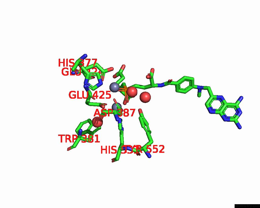
Mono view

Stereo pair view
Mono view
Stereo pair view
A full contact list of Zinc with other atoms in the Zn binding
site number 1 of X-Ray Structure of Human Glutamate Carboxypeptidase II (Gcpii) in Complex with A Transition State Analog of Methotrexate-Glu within 5.0Å range:
|
Zinc binding site 2 out of 2 in 3bi1
Go back to
Zinc binding site 2 out
of 2 in the X-Ray Structure of Human Glutamate Carboxypeptidase II (Gcpii) in Complex with A Transition State Analog of Methotrexate-Glu
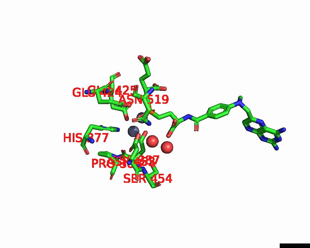
Mono view
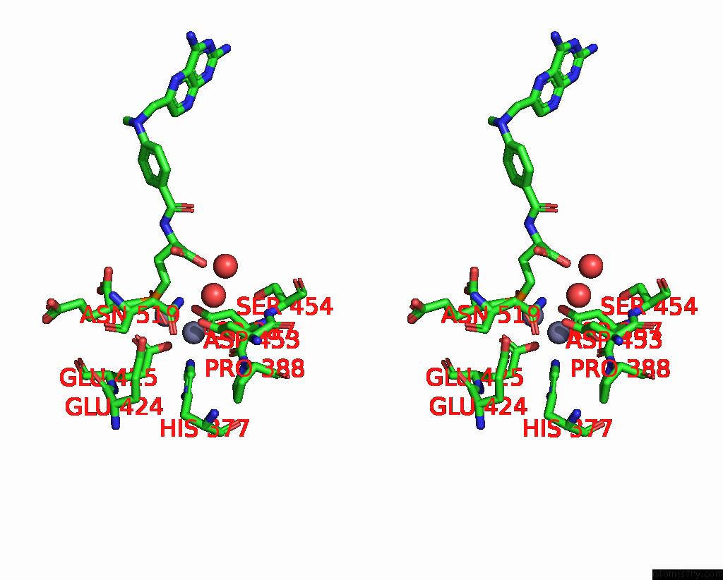
Stereo pair view
Mono view
Stereo pair view
A full contact list of Zinc with other atoms in the Zn binding
site number 2 of X-Ray Structure of Human Glutamate Carboxypeptidase II (Gcpii) in Complex with A Transition State Analog of Methotrexate-Glu within 5.0Å range:
|
Reference:
C.Barinka,
K.Hlouchova,
M.Rovenska,
P.Majer,
M.Dauter,
N.Hin,
Y.S.Ko,
T.Tsukamoto,
B.S.Slusher,
J.Konvalinka,
J.Lubkowski.
Structural Basis of Interactions Between Human Glutamate Carboxypeptidase II and Its Substrate Analogs J.Mol.Biol. V. 376 1438 2008.
ISSN: ISSN 0022-2836
PubMed: 18234225
DOI: 10.1016/J.JMB.2007.12.066
Page generated: Thu Oct 24 11:30:31 2024
ISSN: ISSN 0022-2836
PubMed: 18234225
DOI: 10.1016/J.JMB.2007.12.066
Last articles
Mg in 6YHNMg in 6YHM
Mg in 6YF4
Mg in 6YEM
Mg in 6YE9
Mg in 6YDM
Mg in 6YDL
Mg in 6YDJ
Mg in 6YDK
Mg in 6YCZ