Zinc »
PDB 3ax1-3b7i »
3b37 »
Zinc in PDB 3b37: Crystal Structure of E. Coli Aminopeptidase N in Complex with Tyrosine
Enzymatic activity of Crystal Structure of E. Coli Aminopeptidase N in Complex with Tyrosine
All present enzymatic activity of Crystal Structure of E. Coli Aminopeptidase N in Complex with Tyrosine:
3.4.11.2;
3.4.11.2;
Protein crystallography data
The structure of Crystal Structure of E. Coli Aminopeptidase N in Complex with Tyrosine, PDB code: 3b37
was solved by
A.Addlagatta,
with X-Ray Crystallography technique. A brief refinement statistics is given in the table below:
| Resolution Low / High (Å) | 49.88 / 1.70 |
| Space group | P 31 2 1 |
| Cell size a, b, c (Å), α, β, γ (°) | 120.324, 120.324, 170.345, 90.00, 90.00, 120.00 |
| R / Rfree (%) | 16.2 / 18.4 |
Other elements in 3b37:
The structure of Crystal Structure of E. Coli Aminopeptidase N in Complex with Tyrosine also contains other interesting chemical elements:
| Sodium | (Na) | 2 atoms |
Zinc Binding Sites:
The binding sites of Zinc atom in the Crystal Structure of E. Coli Aminopeptidase N in Complex with Tyrosine
(pdb code 3b37). This binding sites where shown within
5.0 Angstroms radius around Zinc atom.
In total only one binding site of Zinc was determined in the Crystal Structure of E. Coli Aminopeptidase N in Complex with Tyrosine, PDB code: 3b37:
In total only one binding site of Zinc was determined in the Crystal Structure of E. Coli Aminopeptidase N in Complex with Tyrosine, PDB code: 3b37:
Zinc binding site 1 out of 1 in 3b37
Go back to
Zinc binding site 1 out
of 1 in the Crystal Structure of E. Coli Aminopeptidase N in Complex with Tyrosine
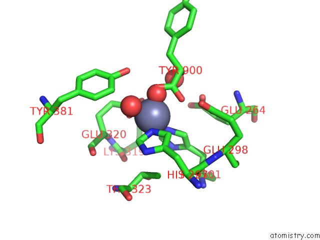
Mono view
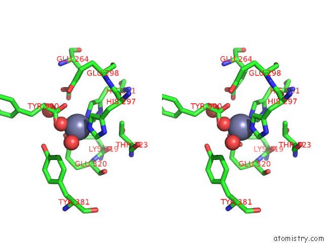
Stereo pair view
Mono view
Stereo pair view
A full contact list of Zinc with other atoms in the Zn binding
site number 1 of Crystal Structure of E. Coli Aminopeptidase N in Complex with Tyrosine within 5.0Å range:
|
Reference:
A.Addlagatta,
L.Gay,
B.W.Matthews.
Structural Basis For the Unusual Specificity of Escherichia Coli Aminopeptidase N. Biochemistry V. 47 5303 2008.
ISSN: ISSN 0006-2960
PubMed: 18416562
DOI: 10.1021/BI7022333
Page generated: Thu Oct 24 11:23:38 2024
ISSN: ISSN 0006-2960
PubMed: 18416562
DOI: 10.1021/BI7022333
Last articles
Mg in 5WRTMg in 5WRJ
Mg in 5WRI
Mg in 5WQA
Mg in 5WQ0
Mg in 5WPM
Mg in 5WPL
Mg in 5WNS
Mg in 5WNQ
Mg in 5WNR