Zinc »
PDB 3adr-3avs »
3aht »
Zinc in PDB 3aht: Crystal Structure of Rice BGLU1 E176Q Mutant in Complex with Laminaribiose
Enzymatic activity of Crystal Structure of Rice BGLU1 E176Q Mutant in Complex with Laminaribiose
All present enzymatic activity of Crystal Structure of Rice BGLU1 E176Q Mutant in Complex with Laminaribiose:
3.2.1.21;
3.2.1.21;
Protein crystallography data
The structure of Crystal Structure of Rice BGLU1 E176Q Mutant in Complex with Laminaribiose, PDB code: 3aht
was solved by
W.Chuenchor,
S.Pengthaisong,
R.C.Robinson,
J.Yuvaniyama,
J.Svasti,
J.R.Ketudat Cairns,
with X-Ray Crystallography technique. A brief refinement statistics is given in the table below:
| Resolution Low / High (Å) | 25.70 / 2.80 |
| Space group | P 21 21 21 |
| Cell size a, b, c (Å), α, β, γ (°) | 79.704, 101.476, 128.291, 90.00, 90.00, 90.00 |
| R / Rfree (%) | 20.5 / 24.8 |
Zinc Binding Sites:
The binding sites of Zinc atom in the Crystal Structure of Rice BGLU1 E176Q Mutant in Complex with Laminaribiose
(pdb code 3aht). This binding sites where shown within
5.0 Angstroms radius around Zinc atom.
In total only one binding site of Zinc was determined in the Crystal Structure of Rice BGLU1 E176Q Mutant in Complex with Laminaribiose, PDB code: 3aht:
In total only one binding site of Zinc was determined in the Crystal Structure of Rice BGLU1 E176Q Mutant in Complex with Laminaribiose, PDB code: 3aht:
Zinc binding site 1 out of 1 in 3aht
Go back to
Zinc binding site 1 out
of 1 in the Crystal Structure of Rice BGLU1 E176Q Mutant in Complex with Laminaribiose
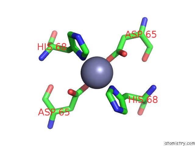
Mono view
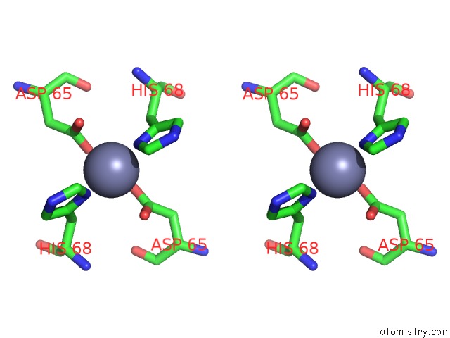
Stereo pair view
Mono view
Stereo pair view
A full contact list of Zinc with other atoms in the Zn binding
site number 1 of Crystal Structure of Rice BGLU1 E176Q Mutant in Complex with Laminaribiose within 5.0Å range:
|
Reference:
W.Chuenchor,
S.Pengthaisong,
R.C.Robinson,
J.Yuvaniyama,
J.Svasti,
J.R.Ketudat Cairns.
The Structural Basis of Oligosaccharide Binding By Rice BGLU1 Beta-Glucosidase J.Struct.Biol. V. 173 169 2011.
ISSN: ISSN 1047-8477
PubMed: 20884352
DOI: 10.1016/J.JSB.2010.09.021
Page generated: Thu Oct 24 11:12:17 2024
ISSN: ISSN 1047-8477
PubMed: 20884352
DOI: 10.1016/J.JSB.2010.09.021
Last articles
Mg in 6YCZMg in 6YCY
Mg in 6YBU
Mg in 6YCX
Mg in 6YB9
Mg in 6YBR
Mg in 6YBO
Mg in 6YBB
Mg in 6YB3
Mg in 6YB5