Zinc »
PDB 2xl9-2xy4 »
2xsn »
Zinc in PDB 2xsn: Crystal Structure of Human Tyrosine Hydroxylase Catalytic Domain
Enzymatic activity of Crystal Structure of Human Tyrosine Hydroxylase Catalytic Domain
All present enzymatic activity of Crystal Structure of Human Tyrosine Hydroxylase Catalytic Domain:
1.14.16.2;
1.14.16.2;
Protein crystallography data
The structure of Crystal Structure of Human Tyrosine Hydroxylase Catalytic Domain, PDB code: 2xsn
was solved by
J.R.C.Muniz,
C.D.O.Cooper,
W.W.Yue,
E.Krysztofinska,
F.Von Delft,
S.Knapp,
O.Gileadi,
C.H.Arrowsmith,
A.M.Edwards,
J.Weigelt,
C.Bountra,
K.L.Kavanagh,
U.Oppermann,
with X-Ray Crystallography technique. A brief refinement statistics is given in the table below:
| Resolution Low / High (Å) | 62.66 / 2.68 |
| Space group | P 65 2 2 |
| Cell size a, b, c (Å), α, β, γ (°) | 191.420, 191.420, 168.160, 90.00, 90.00, 120.00 |
| R / Rfree (%) | 18.9 / 22.2 |
Zinc Binding Sites:
The binding sites of Zinc atom in the Crystal Structure of Human Tyrosine Hydroxylase Catalytic Domain
(pdb code 2xsn). This binding sites where shown within
5.0 Angstroms radius around Zinc atom.
In total 4 binding sites of Zinc where determined in the Crystal Structure of Human Tyrosine Hydroxylase Catalytic Domain, PDB code: 2xsn:
Jump to Zinc binding site number: 1; 2; 3; 4;
In total 4 binding sites of Zinc where determined in the Crystal Structure of Human Tyrosine Hydroxylase Catalytic Domain, PDB code: 2xsn:
Jump to Zinc binding site number: 1; 2; 3; 4;
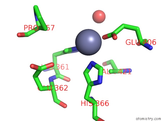
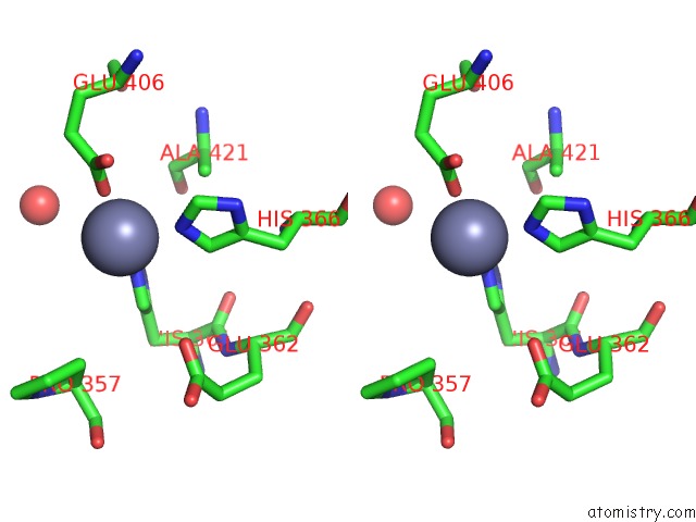
Zinc binding site 1 out of 4 in 2xsn
Go back to
Zinc binding site 1 out
of 4 in the Crystal Structure of Human Tyrosine Hydroxylase Catalytic Domain
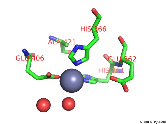
Mono view
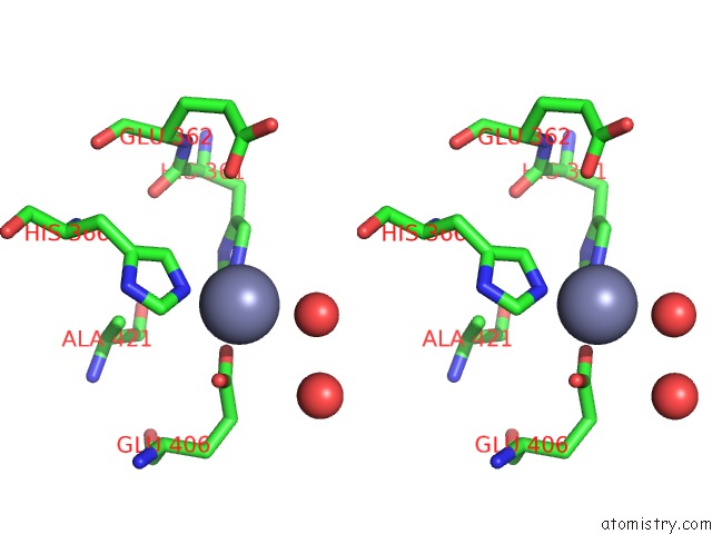
Stereo pair view
Mono view
Stereo pair view
A full contact list of Zinc with other atoms in the Zn binding
site number 1 of Crystal Structure of Human Tyrosine Hydroxylase Catalytic Domain within 5.0Å range:
|
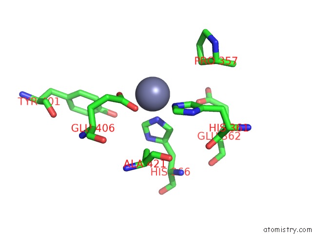
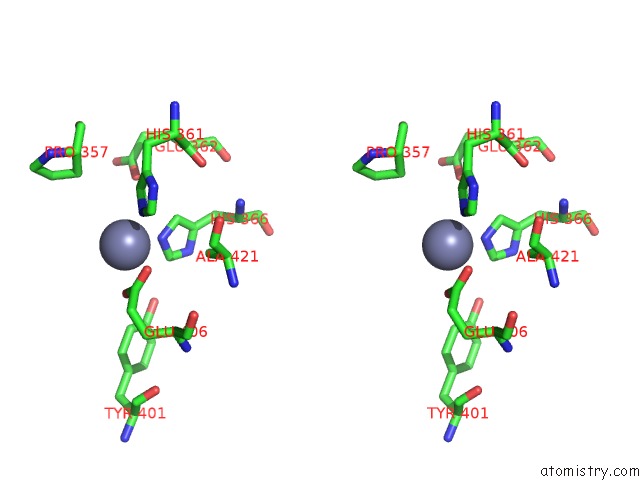
Zinc binding site 2 out of 4 in 2xsn
Go back to
Zinc binding site 2 out
of 4 in the Crystal Structure of Human Tyrosine Hydroxylase Catalytic Domain
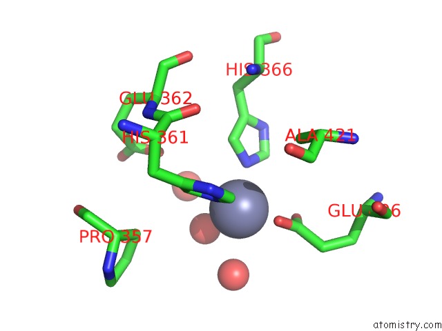
Mono view
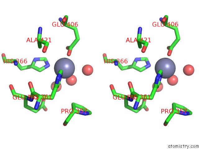
Stereo pair view
Mono view
Stereo pair view
A full contact list of Zinc with other atoms in the Zn binding
site number 2 of Crystal Structure of Human Tyrosine Hydroxylase Catalytic Domain within 5.0Å range:
|
Zinc binding site 3 out of 4 in 2xsn
Go back to
Zinc binding site 3 out
of 4 in the Crystal Structure of Human Tyrosine Hydroxylase Catalytic Domain
Mono view
Stereo pair view
Mono view
Stereo pair view
A full contact list of Zinc with other atoms in the Zn binding
site number 3 of Crystal Structure of Human Tyrosine Hydroxylase Catalytic Domain within 5.0Å range:
|
Zinc binding site 4 out of 4 in 2xsn
Go back to
Zinc binding site 4 out
of 4 in the Crystal Structure of Human Tyrosine Hydroxylase Catalytic Domain
Mono view
Stereo pair view
Mono view
Stereo pair view
A full contact list of Zinc with other atoms in the Zn binding
site number 4 of Crystal Structure of Human Tyrosine Hydroxylase Catalytic Domain within 5.0Å range:
|
Reference:
J.R.C.Muniz,
C.D.O.Cooper,
W.W.Yue,
E.Krysztofinska,
F.Von Delft,
S.Knapp,
O.Gileadi,
C.H.Arrowsmith,
A.M.Edwards,
J.Weigelt,
C.Bountra,
K.L.Kavanagh,
U.Oppermann.
Crystal Structure of Human Tyrosine Hydroxylase Catalytic Domain To Be Published.
Page generated: Wed Aug 20 06:52:33 2025
Last articles
Zn in 3I1UZn in 3HZV
Zn in 3HZM
Zn in 3HUG
Zn in 3HZK
Zn in 3HYP
Zn in 3HYG
Zn in 3HY9
Zn in 3HY7
Zn in 3HXS