Zinc »
PDB 2w22-2wfr »
2wb0 »
Zinc in PDB 2wb0: 2.1 Resolution Structure of the C-Terminal Domain of the Human Adenovirus 5 Ssdna Binding Protein
Protein crystallography data
The structure of 2.1 Resolution Structure of the C-Terminal Domain of the Human Adenovirus 5 Ssdna Binding Protein, PDB code: 2wb0
was solved by
J.Hendle,
P.N.Kanellopoulos,
P.A.Tucker,
with X-Ray Crystallography technique. A brief refinement statistics is given in the table below:
| Resolution Low / High (Å) | 14.95 / 1.95 |
| Space group | P 21 21 21 |
| Cell size a, b, c (Å), α, β, γ (°) | 78.290, 76.660, 64.690, 90.00, 90.00, 90.00 |
| R / Rfree (%) | 18.4 / 24.5 |
Zinc Binding Sites:
The binding sites of Zinc atom in the 2.1 Resolution Structure of the C-Terminal Domain of the Human Adenovirus 5 Ssdna Binding Protein
(pdb code 2wb0). This binding sites where shown within
5.0 Angstroms radius around Zinc atom.
In total 2 binding sites of Zinc where determined in the 2.1 Resolution Structure of the C-Terminal Domain of the Human Adenovirus 5 Ssdna Binding Protein, PDB code: 2wb0:
Jump to Zinc binding site number: 1; 2;
In total 2 binding sites of Zinc where determined in the 2.1 Resolution Structure of the C-Terminal Domain of the Human Adenovirus 5 Ssdna Binding Protein, PDB code: 2wb0:
Jump to Zinc binding site number: 1; 2;
Zinc binding site 1 out of 2 in 2wb0
Go back to
Zinc binding site 1 out
of 2 in the 2.1 Resolution Structure of the C-Terminal Domain of the Human Adenovirus 5 Ssdna Binding Protein
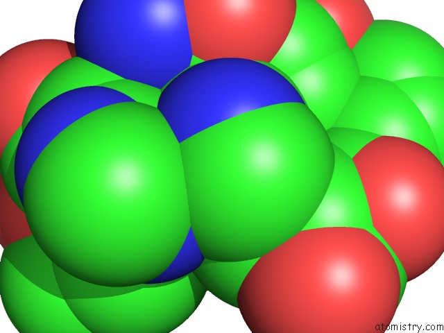
Mono view
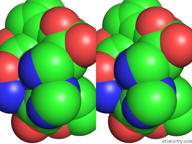
Stereo pair view
Mono view
Stereo pair view
A full contact list of Zinc with other atoms in the Zn binding
site number 1 of 2.1 Resolution Structure of the C-Terminal Domain of the Human Adenovirus 5 Ssdna Binding Protein within 5.0Å range:
|
Zinc binding site 2 out of 2 in 2wb0
Go back to
Zinc binding site 2 out
of 2 in the 2.1 Resolution Structure of the C-Terminal Domain of the Human Adenovirus 5 Ssdna Binding Protein
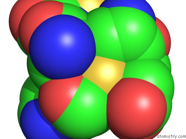
Mono view
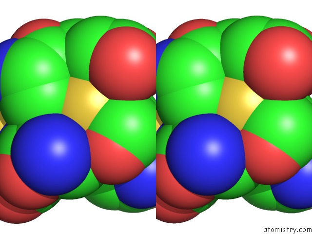
Stereo pair view
Mono view
Stereo pair view
A full contact list of Zinc with other atoms in the Zn binding
site number 2 of 2.1 Resolution Structure of the C-Terminal Domain of the Human Adenovirus 5 Ssdna Binding Protein within 5.0Å range:
|
Reference:
J.Hendle,
P.N.Kanellopoulos,
P.A.Tucker.
High Resolution Structures of the Adenovirus Single-Stranded Dna Binding Protein and the N512P Hinge-Region Mutant To Be Published.
Page generated: Thu Oct 17 04:49:06 2024
Last articles
Fe in 2YXOFe in 2YRS
Fe in 2YXC
Fe in 2YNM
Fe in 2YVJ
Fe in 2YP1
Fe in 2YU2
Fe in 2YU1
Fe in 2YQB
Fe in 2YOO