Zinc »
PDB 2vr6-2w15 »
2vtg »
Zinc in PDB 2vtg: Crystal Structure of Human IBA2, Trigonal Crystal Form
Protein crystallography data
The structure of Crystal Structure of Human IBA2, Trigonal Crystal Form, PDB code: 2vtg
was solved by
J.O.Schulze,
C.Quedenau,
Y.Roske,
A.Turnbull,
U.Mueller,
U.Heinemann,
K.Buessow,
with X-Ray Crystallography technique. A brief refinement statistics is given in the table below:
| Resolution Low / High (Å) | 61.08 / 2.45 |
| Space group | P 32 2 1 |
| Cell size a, b, c (Å), α, β, γ (°) | 70.530, 70.530, 95.190, 90.00, 90.00, 120.00 |
| R / Rfree (%) | 20.4 / 23.2 |
Zinc Binding Sites:
The binding sites of Zinc atom in the Crystal Structure of Human IBA2, Trigonal Crystal Form
(pdb code 2vtg). This binding sites where shown within
5.0 Angstroms radius around Zinc atom.
In total 2 binding sites of Zinc where determined in the Crystal Structure of Human IBA2, Trigonal Crystal Form, PDB code: 2vtg:
Jump to Zinc binding site number: 1; 2;
In total 2 binding sites of Zinc where determined in the Crystal Structure of Human IBA2, Trigonal Crystal Form, PDB code: 2vtg:
Jump to Zinc binding site number: 1; 2;
Zinc binding site 1 out of 2 in 2vtg
Go back to
Zinc binding site 1 out
of 2 in the Crystal Structure of Human IBA2, Trigonal Crystal Form
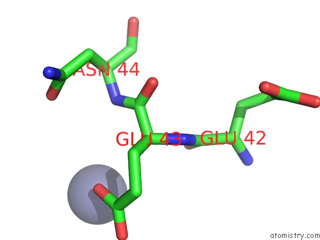
Mono view
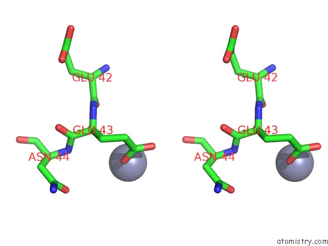
Stereo pair view
Mono view
Stereo pair view
A full contact list of Zinc with other atoms in the Zn binding
site number 1 of Crystal Structure of Human IBA2, Trigonal Crystal Form within 5.0Å range:
|
Zinc binding site 2 out of 2 in 2vtg
Go back to
Zinc binding site 2 out
of 2 in the Crystal Structure of Human IBA2, Trigonal Crystal Form

Mono view
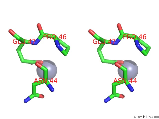
Stereo pair view
Mono view
Stereo pair view
A full contact list of Zinc with other atoms in the Zn binding
site number 2 of Crystal Structure of Human IBA2, Trigonal Crystal Form within 5.0Å range:
|
Reference:
J.O.Schulze,
C.Quedenau,
Y.Roske,
T.Adam,
H.Schuler,
J.Behlke,
A.P.Turnbull,
V.Sievert,
C.Scheich,
U.Mueller,
U.Heinemann,
K.Bussow.
Structural and Functional Characterization of Human Iba Proteins. Febs J. V. 275 4627 2008.
ISSN: ISSN 1742-464X
PubMed: 18699778
DOI: 10.1111/J.1742-4658.2008.06605.X
Page generated: Thu Oct 17 04:33:40 2024
ISSN: ISSN 1742-464X
PubMed: 18699778
DOI: 10.1111/J.1742-4658.2008.06605.X
Last articles
Mg in 9C6KMg in 9C6F
Mg in 9C6C
Mg in 9C67
Mg in 9C59
Mg in 9C5B
Mg in 9C5A
Mg in 9C58
Mg in 9C53
Mg in 9C52