Zinc »
PDB 2qmu-2qyv »
2qn0 »
Zinc in PDB 2qn0: Structure of Botulinum Neurotoxin Serotype C1 Light Chain Protease
Protein crystallography data
The structure of Structure of Botulinum Neurotoxin Serotype C1 Light Chain Protease, PDB code: 2qn0
was solved by
R.Jin,
S.Sikorra,
C.M.Stegmann,
A.Pich,
T.Binz,
A.T.Brunger,
with X-Ray Crystallography technique. A brief refinement statistics is given in the table below:
| Resolution Low / High (Å) | 38.67 / 1.75 |
| Space group | P 31 2 1 |
| Cell size a, b, c (Å), α, β, γ (°) | 107.010, 107.010, 140.390, 90.00, 90.00, 120.00 |
| R / Rfree (%) | 18.8 / 20.5 |
Zinc Binding Sites:
The binding sites of Zinc atom in the Structure of Botulinum Neurotoxin Serotype C1 Light Chain Protease
(pdb code 2qn0). This binding sites where shown within
5.0 Angstroms radius around Zinc atom.
In total only one binding site of Zinc was determined in the Structure of Botulinum Neurotoxin Serotype C1 Light Chain Protease, PDB code: 2qn0:
In total only one binding site of Zinc was determined in the Structure of Botulinum Neurotoxin Serotype C1 Light Chain Protease, PDB code: 2qn0:
Zinc binding site 1 out of 1 in 2qn0
Go back to
Zinc binding site 1 out
of 1 in the Structure of Botulinum Neurotoxin Serotype C1 Light Chain Protease
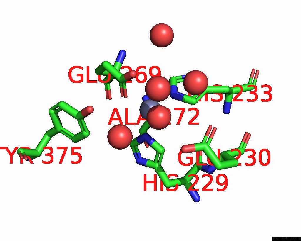
Mono view
Stereo pair view
Mono view
Stereo pair view
A full contact list of Zinc with other atoms in the Zn binding
site number 1 of Structure of Botulinum Neurotoxin Serotype C1 Light Chain Protease within 5.0Å range:
|
Reference:
R.Jin,
S.Sikorra,
C.M.Stegmann,
A.Pich,
T.Binz,
A.T.Brunger.
Structural and Biochemical Studies of Botulinum Neurotoxin Serotype C1 Light Chain Protease: Implications For Dual Substrate Specificity. Biochemistry V. 46 10685 2007.
ISSN: ISSN 0006-2960
PubMed: 17718519
DOI: 10.1021/BI701162D
Page generated: Thu Oct 17 03:31:10 2024
ISSN: ISSN 0006-2960
PubMed: 17718519
DOI: 10.1021/BI701162D
Last articles
Fe in 2YXOFe in 2YRS
Fe in 2YXC
Fe in 2YNM
Fe in 2YVJ
Fe in 2YP1
Fe in 2YU2
Fe in 2YU1
Fe in 2YQB
Fe in 2YOO