Zinc »
PDB 2pou-2q2l »
2px1 »
Zinc in PDB 2px1: Crystal Structure of the Complex of Bovine Lactoferrin C-Lobe with Ribose at 2.5 A Resolution
Protein crystallography data
The structure of Crystal Structure of the Complex of Bovine Lactoferrin C-Lobe with Ribose at 2.5 A Resolution, PDB code: 2px1
was solved by
R.Mir,
G.Vikram,
M.Sinha,
S.Sharma,
P.Kaur,
T.P.Singh,
with X-Ray Crystallography technique. A brief refinement statistics is given in the table below:
| Resolution Low / High (Å) | 19.96 / 2.50 |
| Space group | P 1 21 1 |
| Cell size a, b, c (Å), α, β, γ (°) | 59.906, 49.631, 64.705, 90.00, 105.88, 90.00 |
| R / Rfree (%) | 20.4 / 22.8 |
Other elements in 2px1:
The structure of Crystal Structure of the Complex of Bovine Lactoferrin C-Lobe with Ribose at 2.5 A Resolution also contains other interesting chemical elements:
| Iron | (Fe) | 1 atom |
Zinc Binding Sites:
The binding sites of Zinc atom in the Crystal Structure of the Complex of Bovine Lactoferrin C-Lobe with Ribose at 2.5 A Resolution
(pdb code 2px1). This binding sites where shown within
5.0 Angstroms radius around Zinc atom.
In total 2 binding sites of Zinc where determined in the Crystal Structure of the Complex of Bovine Lactoferrin C-Lobe with Ribose at 2.5 A Resolution, PDB code: 2px1:
Jump to Zinc binding site number: 1; 2;
In total 2 binding sites of Zinc where determined in the Crystal Structure of the Complex of Bovine Lactoferrin C-Lobe with Ribose at 2.5 A Resolution, PDB code: 2px1:
Jump to Zinc binding site number: 1; 2;
Zinc binding site 1 out of 2 in 2px1
Go back to
Zinc binding site 1 out
of 2 in the Crystal Structure of the Complex of Bovine Lactoferrin C-Lobe with Ribose at 2.5 A Resolution
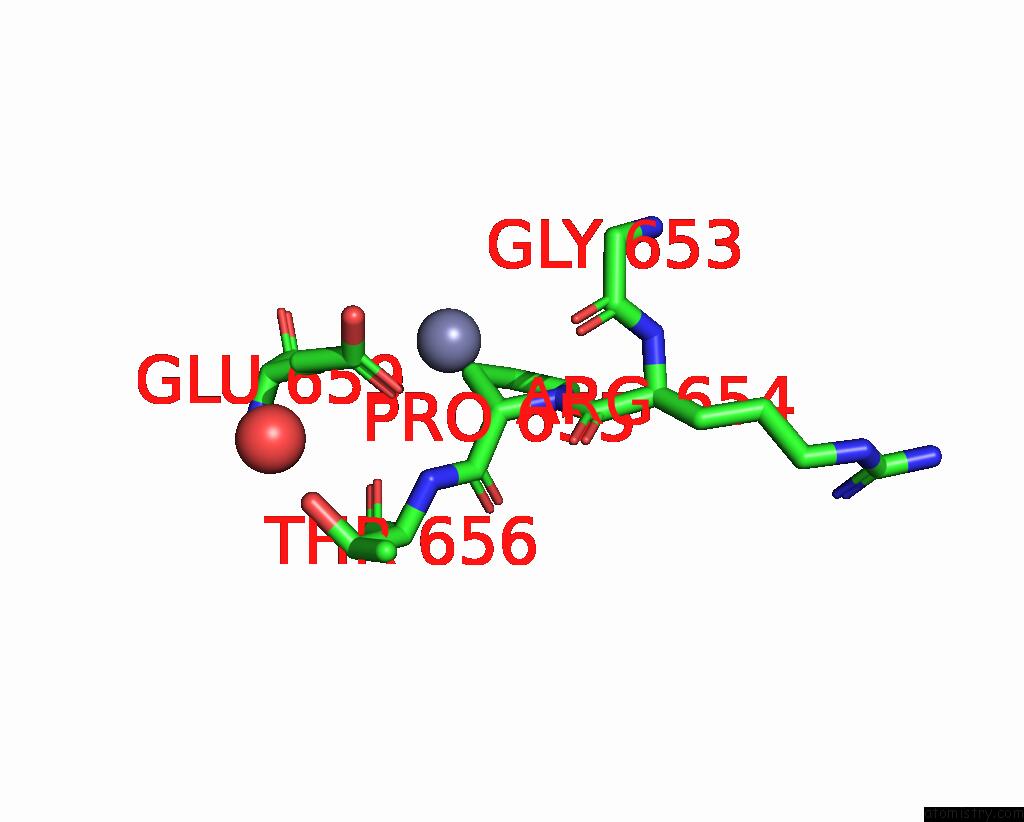
Mono view
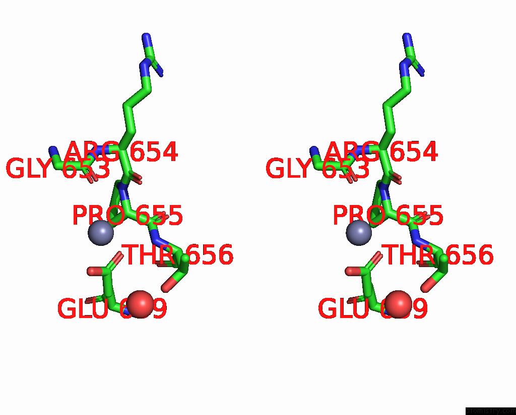
Stereo pair view
Mono view
Stereo pair view
A full contact list of Zinc with other atoms in the Zn binding
site number 1 of Crystal Structure of the Complex of Bovine Lactoferrin C-Lobe with Ribose at 2.5 A Resolution within 5.0Å range:
|
Zinc binding site 2 out of 2 in 2px1
Go back to
Zinc binding site 2 out
of 2 in the Crystal Structure of the Complex of Bovine Lactoferrin C-Lobe with Ribose at 2.5 A Resolution
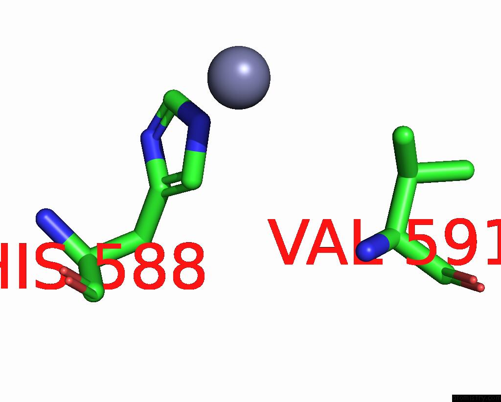
Mono view
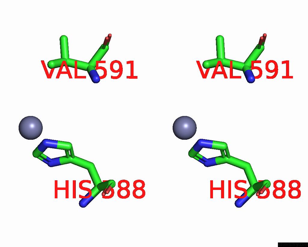
Stereo pair view
Mono view
Stereo pair view
A full contact list of Zinc with other atoms in the Zn binding
site number 2 of Crystal Structure of the Complex of Bovine Lactoferrin C-Lobe with Ribose at 2.5 A Resolution within 5.0Å range:
|
Reference:
R.Mir,
G.Vikram,
M.Sinha,
N.Singh,
S.Sharma,
P.Kaur,
M.Perbandt,
C.Betzel,
T.P.Singh.
Crystal Structure of the Complex of Bovine Lactoferrin C-Lobe with Ribose at 2.5 A Resolution To Be Published.
Page generated: Thu Oct 17 03:17:41 2024
Last articles
Fe in 8PWYFe in 8PXQ
Fe in 8PY5
Fe in 8PWS
Fe in 8PXP
Fe in 8PP4
Fe in 8PUR
Fe in 8PVD
Fe in 8PU0
Fe in 8PVC