Zinc »
PDB 2pou-2q2l »
2pu0 »
Zinc in PDB 2pu0: Crystal Structure of the T. Brucei Enolase Complexed with Phosphonoacetohydroxamate (Pah), HIS156-in Conformation
Enzymatic activity of Crystal Structure of the T. Brucei Enolase Complexed with Phosphonoacetohydroxamate (Pah), HIS156-in Conformation
All present enzymatic activity of Crystal Structure of the T. Brucei Enolase Complexed with Phosphonoacetohydroxamate (Pah), HIS156-in Conformation:
4.2.1.11;
4.2.1.11;
Protein crystallography data
The structure of Crystal Structure of the T. Brucei Enolase Complexed with Phosphonoacetohydroxamate (Pah), HIS156-in Conformation, PDB code: 2pu0
was solved by
M.V.A.S.Navarro,
D.J.Rigden,
R.C.Garratt,
S.M.G.Dias,
with X-Ray Crystallography technique. A brief refinement statistics is given in the table below:
| Resolution Low / High (Å) | 17.72 / 1.90 |
| Space group | C 2 2 21 |
| Cell size a, b, c (Å), α, β, γ (°) | 74.946, 110.760, 109.219, 90.00, 90.00, 90.00 |
| R / Rfree (%) | 16.5 / 20.5 |
Zinc Binding Sites:
The binding sites of Zinc atom in the Crystal Structure of the T. Brucei Enolase Complexed with Phosphonoacetohydroxamate (Pah), HIS156-in Conformation
(pdb code 2pu0). This binding sites where shown within
5.0 Angstroms radius around Zinc atom.
In total 3 binding sites of Zinc where determined in the Crystal Structure of the T. Brucei Enolase Complexed with Phosphonoacetohydroxamate (Pah), HIS156-in Conformation, PDB code: 2pu0:
Jump to Zinc binding site number: 1; 2; 3;
In total 3 binding sites of Zinc where determined in the Crystal Structure of the T. Brucei Enolase Complexed with Phosphonoacetohydroxamate (Pah), HIS156-in Conformation, PDB code: 2pu0:
Jump to Zinc binding site number: 1; 2; 3;
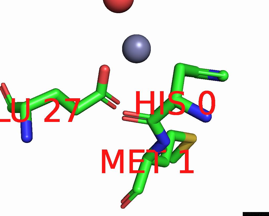
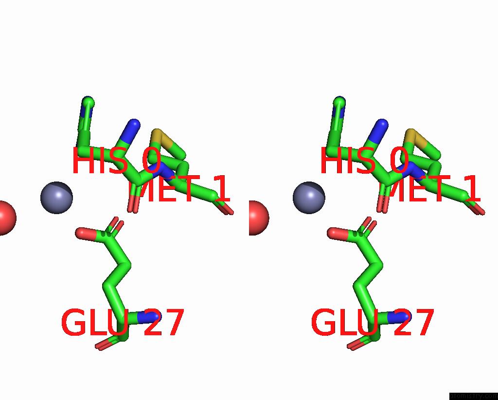
Zinc binding site 1 out of 3 in 2pu0
Go back to
Zinc binding site 1 out
of 3 in the Crystal Structure of the T. Brucei Enolase Complexed with Phosphonoacetohydroxamate (Pah), HIS156-in Conformation
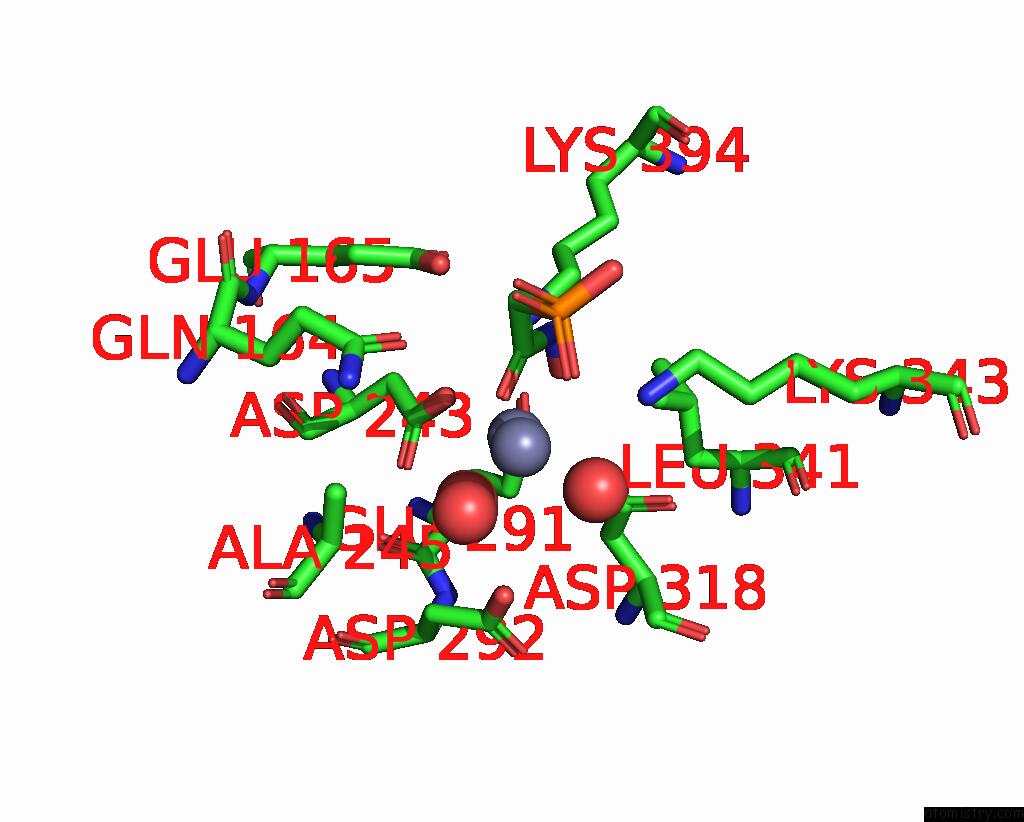
Mono view
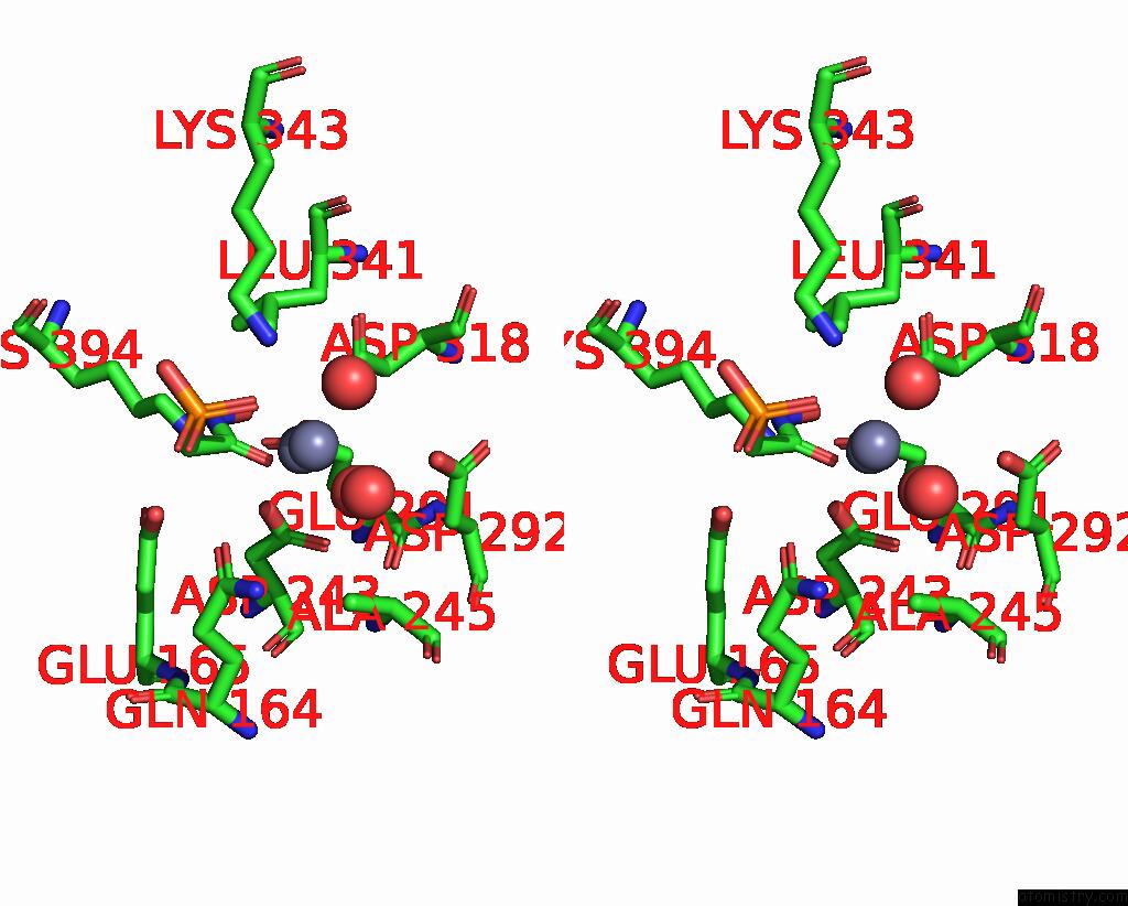
Stereo pair view
Mono view
Stereo pair view
A full contact list of Zinc with other atoms in the Zn binding
site number 1 of Crystal Structure of the T. Brucei Enolase Complexed with Phosphonoacetohydroxamate (Pah), HIS156-in Conformation within 5.0Å range:
|
Zinc binding site 2 out of 3 in 2pu0
Go back to
Zinc binding site 2 out
of 3 in the Crystal Structure of the T. Brucei Enolase Complexed with Phosphonoacetohydroxamate (Pah), HIS156-in Conformation
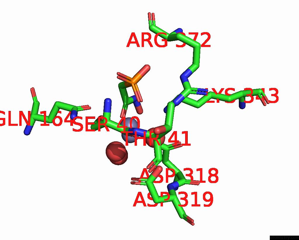
Mono view
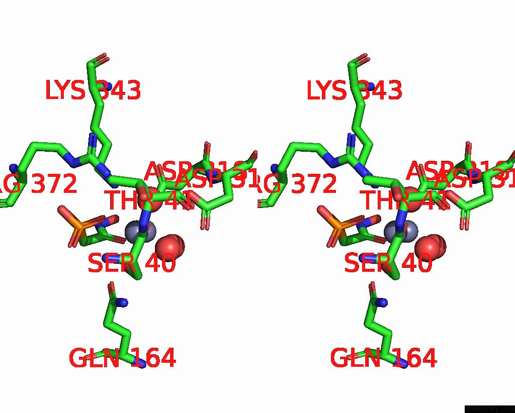
Stereo pair view
Mono view
Stereo pair view
A full contact list of Zinc with other atoms in the Zn binding
site number 2 of Crystal Structure of the T. Brucei Enolase Complexed with Phosphonoacetohydroxamate (Pah), HIS156-in Conformation within 5.0Å range:
|
Zinc binding site 3 out of 3 in 2pu0
Go back to
Zinc binding site 3 out
of 3 in the Crystal Structure of the T. Brucei Enolase Complexed with Phosphonoacetohydroxamate (Pah), HIS156-in Conformation
Mono view
Stereo pair view
Mono view
Stereo pair view
A full contact list of Zinc with other atoms in the Zn binding
site number 3 of Crystal Structure of the T. Brucei Enolase Complexed with Phosphonoacetohydroxamate (Pah), HIS156-in Conformation within 5.0Å range:
|
Reference:
M.V.Navarro,
S.M.Gomes Dias,
L.V.Mello,
M.T.Da Silva Giotto,
S.Gavalda,
C.Blonski,
R.C.Garratt,
D.J.Rigden.
Structural Flexibility in Trypanosoma Brucei Enolase Revealed By X-Ray Crystallography and Molecular Dynamics. Febs J. V. 274 5077 2007.
ISSN: ISSN 1742-464X
PubMed: 17822439
DOI: 10.1111/J.1742-4658.2007.06027.X
Page generated: Thu Oct 17 03:13:24 2024
ISSN: ISSN 1742-464X
PubMed: 17822439
DOI: 10.1111/J.1742-4658.2007.06027.X
Last articles
Fe in 8P8GFe in 8POZ
Fe in 8POX
Fe in 8POY
Fe in 8POW
Fe in 8POU
Fe in 8POV
Fe in 8PMK
Fe in 8PNV
Fe in 8PNU