Atomistry » Zinc » PDB 2pcx-2pot » 2ped
Atomistry »
  Zinc »
    PDB 2pcx-2pot »
      2ped »

Zinc in PDB 2ped: Crystallographic Model of 9-Cis-Rhodopsin

Protein crystallography data

The structure of Crystallographic Model of 9-Cis-Rhodopsin, PDB code: 2ped was solved by H.Nakamichi, T.Okada, with X-Ray Crystallography technique. A brief refinement statistics is given in the table below:

Resolution Low / High (Å) 50.00 / 2.95
Space group P 41
Cell size a, b, c (Å), α, β, γ (°) 95.960, 95.960, 150.840, 90.00, 90.00, 90.00
R / Rfree (%) 24.3 / 28.9

Other elements in 2ped:

The structure of Crystallographic Model of 9-Cis-Rhodopsin also contains other interesting chemical elements:

Mercury (Hg) 6 atoms

Zinc Binding Sites:

The binding sites of Zinc atom in the Crystallographic Model of 9-Cis-Rhodopsin (pdb code 2ped). This binding sites where shown within 5.0 Angstroms radius around Zinc atom.
In total 7 binding sites of Zinc where determined in the Crystallographic Model of 9-Cis-Rhodopsin, PDB code: 2ped:
Jump to Zinc binding site number: 1; 2; 3; 4; 5; 6; 7;

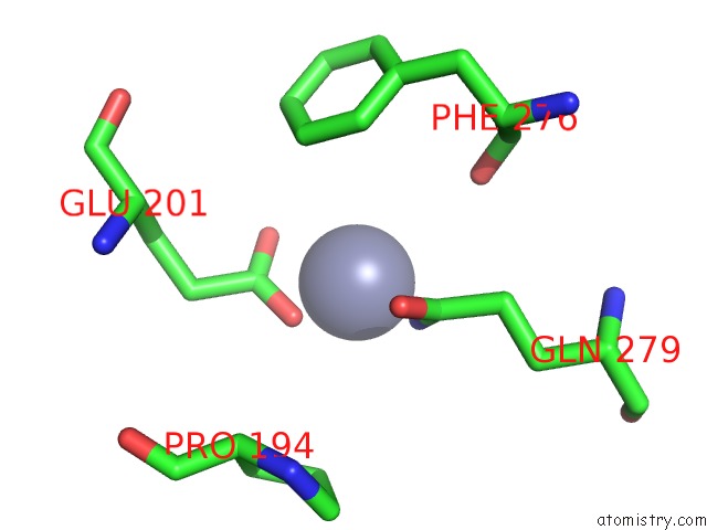
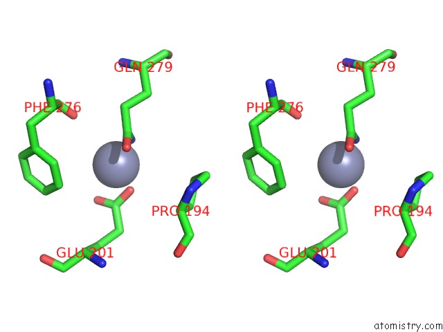
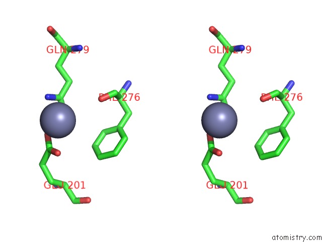
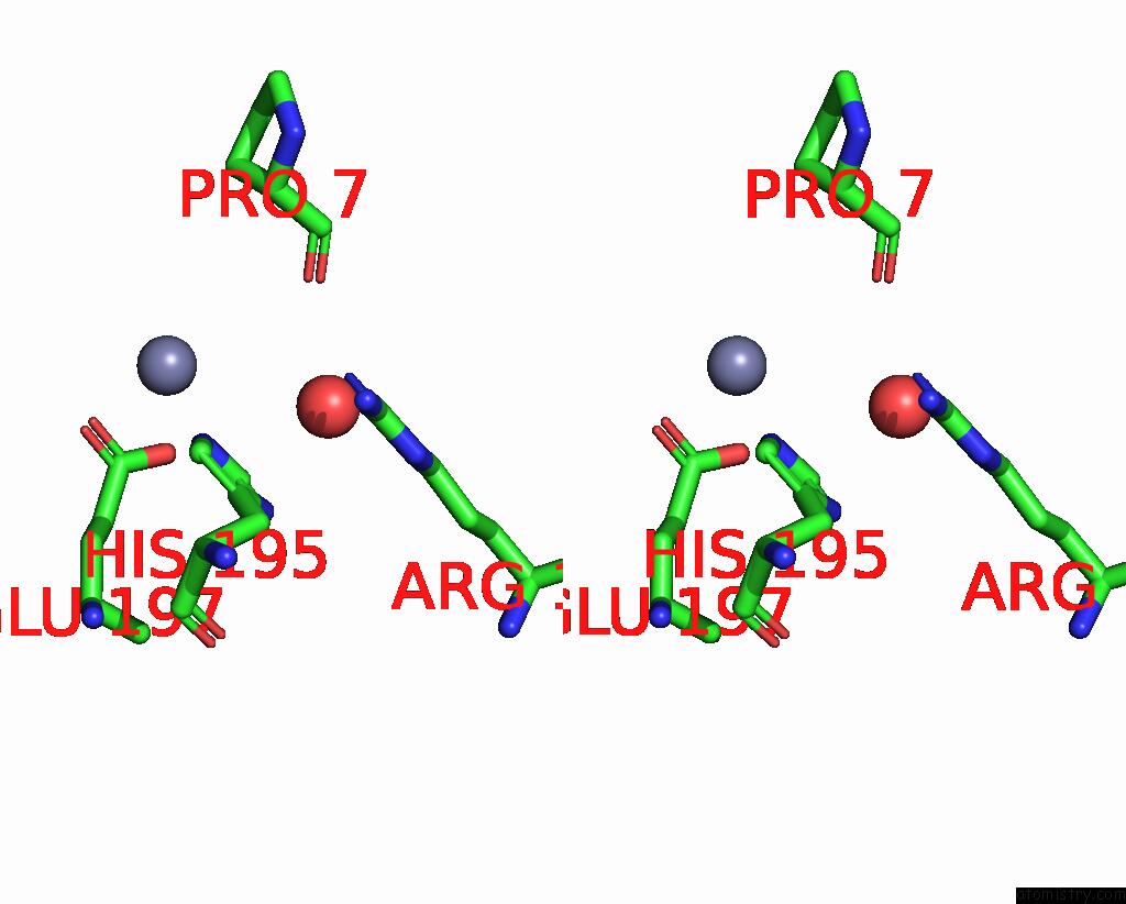
Zinc binding site 1 out of 7 in 2ped

Go back to Zinc Binding Sites List in 2ped
Zinc binding site 1 out of 7 in the Crystallographic Model of 9-Cis-Rhodopsin


Mono view


Stereo pair view

A full contact list of Zinc with other atoms in the Zn binding site number 1 of Crystallographic Model of 9-Cis-Rhodopsin within 5.0Å range:
probe atom residue distance (Å) B Occ
B:Zn956

b:68.2
occ:0.73
NE2 B:HIS195 2.3 65.8 1.0
OE1 B:GLU197 2.7 79.2 1.0
CD2 B:HIS195 3.0 66.2 1.0
CD B:GLU197 3.3 76.8 1.0
OE2 B:GLU197 3.3 75.3 1.0
CE1 B:HIS195 3.4 66.0 1.0
O B:PRO7 3.8 56.8 1.0
CG B:HIS195 4.3 65.5 1.0
ND1 B:HIS195 4.4 66.4 1.0
NH1 B:ARG177 4.6 49.6 1.0
CG B:GLU197 4.6 73.4 1.0
O B:HOH2103 4.7 45.9 1.0
C B:PRO7 4.8 56.2 1.0
CB B:GLU197 4.8 69.6 1.0
CB B:PRO7 4.9 55.1 1.0
CA B:PRO7 4.9 55.5 1.0

Zinc binding site 2 out of 7 in 2ped

Go back to Zinc Binding Sites List in 2ped
Zinc binding site 2 out of 7 in the Crystallographic Model of 9-Cis-Rhodopsin


Mono view


Stereo pair view

A full contact list of Zinc with other atoms in the Zn binding site number 2 of Crystallographic Model of 9-Cis-Rhodopsin within 5.0Å range:
probe atom residue distance (Å) B Occ
A:Zn957

b:53.2
occ:0.69
NE2 A:GLN279 1.9 61.0 1.0
OE2 A:GLU201 2.0 58.3 1.0
OE1 A:GLU201 2.4 60.4 1.0
CD A:GLU201 2.5 59.4 1.0
CD A:GLN279 2.8 60.7 1.0
OE1 A:GLN279 3.2 62.0 1.0
CG A:GLU201 4.0 59.6 1.0
O A:PHE276 4.0 54.8 1.0
CG A:GLN279 4.1 58.2 1.0
CE1 A:PHE276 4.4 51.2 1.0
CZ A:PHE276 4.6 50.4 1.0
CG A:PRO194 4.8 61.5 1.0
CD1 A:PHE276 4.9 50.3 1.0
CB A:GLU201 5.0 58.5 1.0
CB A:GLN279 5.0 54.2 1.0

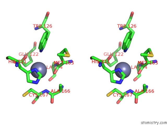
Zinc binding site 3 out of 7 in 2ped

Go back to Zinc Binding Sites List in 2ped
Zinc binding site 3 out of 7 in the Crystallographic Model of 9-Cis-Rhodopsin


Mono view


Stereo pair view

A full contact list of Zinc with other atoms in the Zn binding site number 3 of Crystallographic Model of 9-Cis-Rhodopsin within 5.0Å range:
probe atom residue distance (Å) B Occ
B:Zn958

b:41.7
occ:0.55
OE2 B:GLU201 2.0 64.0 1.0
NE2 B:GLN279 2.4 51.9 1.0
OE1 B:GLU201 2.5 65.0 1.0
CD B:GLU201 2.6 65.9 1.0
CD B:GLN279 2.9 53.4 1.0
OE1 B:GLN279 3.0 53.8 1.0
O B:PHE276 3.8 59.2 1.0
CG B:GLN279 4.1 53.1 1.0
CG B:GLU201 4.1 66.1 1.0
CE1 B:PHE276 4.3 57.7 1.0
CZ B:PHE276 4.5 56.9 1.0
CD1 B:PHE276 4.7 57.9 1.0
CB B:GLN279 4.9 53.5 1.0
C B:PHE276 5.0 58.3 1.0

Zinc binding site 4 out of 7 in 2ped

Go back to Zinc Binding Sites List in 2ped
Zinc binding site 4 out of 7 in the Crystallographic Model of 9-Cis-Rhodopsin


Mono view


Stereo pair view

A full contact list of Zinc with other atoms in the Zn binding site number 4 of Crystallographic Model of 9-Cis-Rhodopsin within 5.0Å range:
probe atom residue distance (Å) B Occ
A:Zn959

b:85.3
occ:0.86
NZ A:LYS311 2.1 71.5 1.0
N A:ASP330 2.2 0.4 1.0
CE1 B:HIS100 2.3 57.5 1.0
C A:GLY329 2.6 1.0 1.0
CB A:ASP330 2.6 0.3 1.0
CA A:ASP330 2.9 0.9 1.0
CA A:GLY329 3.0 0.9 1.0
NE2 B:HIS100 3.0 58.8 1.0
CG A:ASP330 3.1 0.9 1.0
CE A:LYS311 3.3 71.6 1.0
OD2 A:ASP330 3.3 0.5 1.0
O A:GLY329 3.4 0.5 1.0
ND1 B:HIS100 3.5 56.1 1.0
C A:ASP330 3.8 0.9 1.0
OD1 A:ASP330 3.9 0.7 1.0
N A:ASP331 4.2 0.6 1.0
CD2 B:HIS100 4.3 56.9 1.0
N A:GLY329 4.5 0.8 1.0
CG B:HIS100 4.5 55.3 1.0
O A:ASP330 4.6 0.5 1.0
CD A:LYS311 4.6 70.0 1.0
O A:ASP331 4.7 0.2 1.0
NH2 A:ARG314 4.8 52.4 1.0
OH B:TYR96 4.9 62.7 1.0
CE1 B:TYR96 4.9 61.3 1.0
O A:LEU328 4.9 0.2 1.0

Zinc binding site 5 out of 7 in 2ped

Go back to Zinc Binding Sites List in 2ped
Zinc binding site 5 out of 7 in the Crystallographic Model of 9-Cis-Rhodopsin


Mono view


Stereo pair view

A full contact list of Zinc with other atoms in the Zn binding site number 5 of Crystallographic Model of 9-Cis-Rhodopsin within 5.0Å range:
probe atom residue distance (Å) B Occ
A:Zn962

b:0.7
occ:0.60
ND1 A:HIS211 2.5 62.0 1.0
NE1 A:TRP126 2.7 64.1 1.0
OE1 A:GLU122 2.7 52.6 1.0
O A:MET163 2.8 60.5 1.0
CE1 A:HIS211 2.8 62.6 1.0
CG A:HIS211 3.0 60.5 1.0
C A:MET163 3.3 60.1 1.0
CD A:GLU122 3.4 52.5 1.0
CD1 A:TRP126 3.4 63.6 1.0
NE2 A:HIS211 3.5 63.1 1.0
CG A:GLU122 3.5 52.0 1.0
CD2 A:HIS211 3.6 62.4 1.0
CB A:HIS211 3.7 58.1 1.0
CA A:ALA164 3.7 59.1 1.0
N A:ALA164 3.7 59.8 1.0
CB A:GLU122 3.8 52.3 1.0
CE2 A:TRP126 3.9 62.5 1.0
CB A:CYS167 4.0 59.3 1.0
CB A:MET163 4.2 60.5 1.0
CA A:MET163 4.3 60.3 1.0
N A:CYS167 4.3 59.9 1.0
CZ2 A:TRP126 4.4 61.8 1.0
OE2 A:GLU122 4.5 53.3 1.0
C A:ALA164 4.5 58.4 1.0
O A:ALA164 4.5 59.2 1.0
CG A:TRP126 4.7 62.0 1.0
CA A:HIS211 4.7 57.6 1.0
CA A:CYS167 4.7 60.2 1.0
CB A:ALA164 4.9 60.4 1.0
CA A:GLU122 4.9 52.6 1.0
CD2 A:TRP126 4.9 62.1 1.0

Zinc binding site 6 out of 7 in 2ped

Go back to Zinc Binding Sites List in 2ped
Zinc binding site 6 out of 7 in the Crystallographic Model of 9-Cis-Rhodopsin


Mono view


Stereo pair view

A full contact list of Zinc with other atoms in the Zn binding site number 6 of Crystallographic Model of 9-Cis-Rhodopsin within 5.0Å range:
probe atom residue distance (Å) B Occ
B:Zn963

b:0.2
occ:0.64
ND1 B:HIS211 2.5 75.2 1.0
O B:MET163 2.8 69.6 1.0
CE1 B:HIS211 2.8 75.7 1.0
CG B:HIS211 2.8 73.3 1.0
NE1 B:TRP126 3.3 76.8 1.0
OE1 B:GLU122 3.3 63.1 1.0
NE2 B:HIS211 3.3 75.1 1.0
CD2 B:HIS211 3.3 74.8 1.0
C B:MET163 3.4 69.5 1.0
CB B:HIS211 3.5 70.7 1.0
CB B:CYS167 3.5 59.0 1.0
CD B:GLU122 3.7 61.7 1.0
N B:CYS167 3.7 61.1 1.0
CG B:GLU122 3.7 60.6 1.0
CA B:ALA164 3.9 65.6 1.0
N B:ALA164 3.9 67.8 1.0
O B:ALA164 4.0 64.9 1.0
CB B:GLU122 4.1 59.9 1.0
CD1 B:TRP126 4.1 76.8 1.0
CA B:CYS167 4.2 60.0 1.0
CE2 B:TRP126 4.3 77.3 1.0
C B:ALA164 4.3 64.4 1.0
CA B:MET163 4.4 70.4 1.0
CB B:MET163 4.4 71.8 1.0
CB B:ALA166 4.6 62.0 1.0
OE2 B:GLU122 4.6 62.0 1.0
CZ2 B:TRP126 4.6 77.9 1.0
CA B:HIS211 4.6 68.9 1.0
C B:ALA166 4.7 62.0 1.0
SG B:CYS167 4.9 55.2 1.0

Zinc binding site 7 out of 7 in 2ped

Go back to Zinc Binding Sites List in 2ped
Zinc binding site 7 out of 7 in the Crystallographic Model of 9-Cis-Rhodopsin


Mono view


Stereo pair view

A full contact list of Zinc with other atoms in the Zn binding site number 7 of Crystallographic Model of 9-Cis-Rhodopsin within 5.0Å range:
probe atom residue distance (Å) B Occ
A:Zn2011

b:53.0
occ:0.71
NE2 A:HIS195 2.3 60.9 1.0
OE1 A:GLU197 2.6 79.6 1.0
OE2 A:GLU197 3.0 77.7 1.0
CD A:GLU197 3.0 78.1 1.0
CD2 A:HIS195 3.2 60.7 1.0
CE1 A:HIS195 3.3 60.9 1.0
O A:PRO7 4.1 48.5 1.0
O A:HOH2062 4.3 55.0 1.0
ND1 A:HIS195 4.4 60.7 1.0
CG A:GLU197 4.4 74.3 1.0
CG A:HIS195 4.4 61.3 1.0
NH1 A:ARG177 4.5 51.2 1.0
CB A:GLU197 4.6 68.9 1.0
C A:PRO7 5.0 48.8 1.0

Reference:

H.Nakamichi, V.Buss, T.Okada. Photoisomerization Mechanism of Rhodopsin and 9-Cis-Rhodopsin Revealed By X-Ray Crystallography Biophys.J. V. 92 L106 2007.
ISSN: ISSN 0006-3495
PubMed: 17449675
DOI: 10.1529/BIOPHYSJ.107.108225
Page generated: Thu Oct 17 02:59:36 2024

Last articles

Fe in 2YXO
Fe in 2YRS
Fe in 2YXC
Fe in 2YNM
Fe in 2YVJ
Fe in 2YP1
Fe in 2YU2
Fe in 2YU1
Fe in 2YQB
Fe in 2YOO
© Copyright 2008-2020 by atomistry.com
Home   |    Site Map   |    Copyright   |    Contact us   |    Privacy