Zinc »
PDB 2o8h-2omh »
2ofk »
Zinc in PDB 2ofk: Crystal Structure of 3-Methyladenine Dna Glycosylase I (Tag)
Protein crystallography data
The structure of Crystal Structure of 3-Methyladenine Dna Glycosylase I (Tag), PDB code: 2ofk
was solved by
A.H.Metz,
T.Hollis,
B.F.Eichman,
with X-Ray Crystallography technique. A brief refinement statistics is given in the table below:
| Resolution Low / High (Å) | 50.00 / 1.50 |
| Space group | P 1 21 1 |
| Cell size a, b, c (Å), α, β, γ (°) | 57.455, 63.681, 62.127, 90.00, 106.94, 90.00 |
| R / Rfree (%) | 16.1 / 19.6 |
Zinc Binding Sites:
The binding sites of Zinc atom in the Crystal Structure of 3-Methyladenine Dna Glycosylase I (Tag)
(pdb code 2ofk). This binding sites where shown within
5.0 Angstroms radius around Zinc atom.
In total 2 binding sites of Zinc where determined in the Crystal Structure of 3-Methyladenine Dna Glycosylase I (Tag), PDB code: 2ofk:
Jump to Zinc binding site number: 1; 2;
In total 2 binding sites of Zinc where determined in the Crystal Structure of 3-Methyladenine Dna Glycosylase I (Tag), PDB code: 2ofk:
Jump to Zinc binding site number: 1; 2;
Zinc binding site 1 out of 2 in 2ofk
Go back to
Zinc binding site 1 out
of 2 in the Crystal Structure of 3-Methyladenine Dna Glycosylase I (Tag)
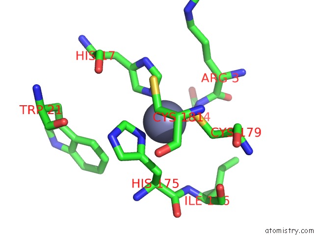
Mono view
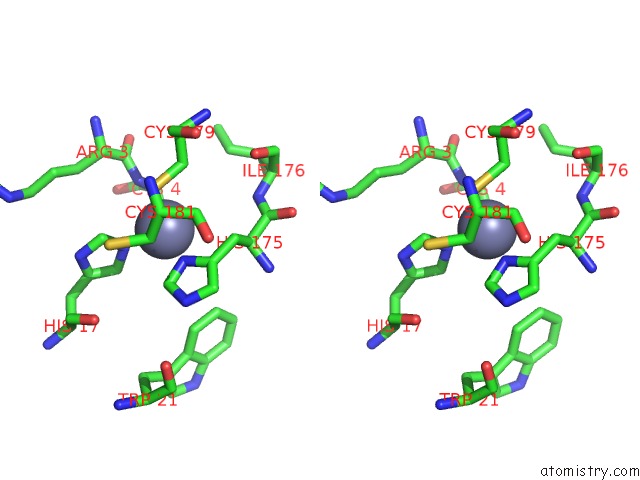
Stereo pair view
Mono view
Stereo pair view
A full contact list of Zinc with other atoms in the Zn binding
site number 1 of Crystal Structure of 3-Methyladenine Dna Glycosylase I (Tag) within 5.0Å range:
|
Zinc binding site 2 out of 2 in 2ofk
Go back to
Zinc binding site 2 out
of 2 in the Crystal Structure of 3-Methyladenine Dna Glycosylase I (Tag)
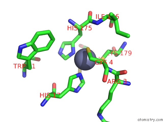
Mono view
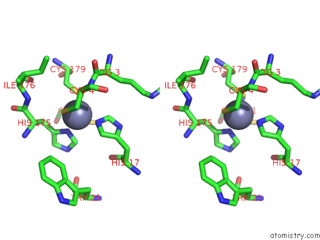
Stereo pair view
Mono view
Stereo pair view
A full contact list of Zinc with other atoms in the Zn binding
site number 2 of Crystal Structure of 3-Methyladenine Dna Glycosylase I (Tag) within 5.0Å range:
|
Reference:
A.H.Metz,
T.Hollis,
B.F.Eichman.
Dna Damage Recognition and Repair By 3-Methyladenine Dna Glycosylase I (Tag). Embo J. V. 26 2411 2007.
ISSN: ISSN 0261-4189
PubMed: 17410210
DOI: 10.1038/SJ.EMBOJ.7601649
Page generated: Thu Oct 17 02:37:35 2024
ISSN: ISSN 0261-4189
PubMed: 17410210
DOI: 10.1038/SJ.EMBOJ.7601649
Last articles
Fe in 2YXOFe in 2YRS
Fe in 2YXC
Fe in 2YNM
Fe in 2YVJ
Fe in 2YP1
Fe in 2YU2
Fe in 2YU1
Fe in 2YQB
Fe in 2YOO