Zinc »
PDB 2o8h-2omh »
2o8h »
Zinc in PDB 2o8h: Crystal Structure of the Catalytic Domain of Rat Phosphodiesterase 10A
Enzymatic activity of Crystal Structure of the Catalytic Domain of Rat Phosphodiesterase 10A
All present enzymatic activity of Crystal Structure of the Catalytic Domain of Rat Phosphodiesterase 10A:
3.1.4.17;
3.1.4.17;
Protein crystallography data
The structure of Crystal Structure of the Catalytic Domain of Rat Phosphodiesterase 10A, PDB code: 2o8h
was solved by
J.Pandit,
E.S.Marr,
with X-Ray Crystallography technique. A brief refinement statistics is given in the table below:
| Resolution Low / High (Å) | 30.00 / 1.80 |
| Space group | H 3 |
| Cell size a, b, c (Å), α, β, γ (°) | 120.622, 120.622, 82.143, 90.00, 90.00, 120.00 |
| R / Rfree (%) | 22.4 / 27.3 |
Other elements in 2o8h:
The structure of Crystal Structure of the Catalytic Domain of Rat Phosphodiesterase 10A also contains other interesting chemical elements:
| Magnesium | (Mg) | 1 atom |
Zinc Binding Sites:
The binding sites of Zinc atom in the Crystal Structure of the Catalytic Domain of Rat Phosphodiesterase 10A
(pdb code 2o8h). This binding sites where shown within
5.0 Angstroms radius around Zinc atom.
In total only one binding site of Zinc was determined in the Crystal Structure of the Catalytic Domain of Rat Phosphodiesterase 10A, PDB code: 2o8h:
In total only one binding site of Zinc was determined in the Crystal Structure of the Catalytic Domain of Rat Phosphodiesterase 10A, PDB code: 2o8h:
Zinc binding site 1 out of 1 in 2o8h
Go back to
Zinc binding site 1 out
of 1 in the Crystal Structure of the Catalytic Domain of Rat Phosphodiesterase 10A
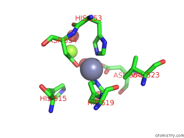
Mono view
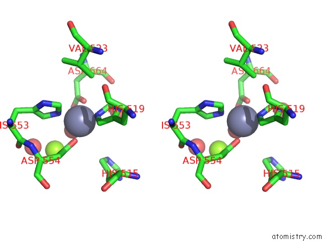
Stereo pair view
Mono view
Stereo pair view
A full contact list of Zinc with other atoms in the Zn binding
site number 1 of Crystal Structure of the Catalytic Domain of Rat Phosphodiesterase 10A within 5.0Å range:
|
Reference:
T.A.Chappie,
J.M.Humphrey,
M.P.Allen,
K.G.Estep,
C.B.Fox,
L.A.Lebel,
S.Liras,
E.S.Marr,
F.S.Menniti,
J.Pandit,
C.J.Schmidt,
M.Tu,
R.D.Williams,
F.V.Yang.
Discovery of A Series of 6,7-Dimethoxy-4-Pyrrolidylquinazoline PDE10A Inhibitors J.Med.Chem. V. 50 182 2007.
ISSN: ISSN 0022-2623
PubMed: 17228859
DOI: 10.1021/JM060653B
Page generated: Thu Oct 17 02:31:35 2024
ISSN: ISSN 0022-2623
PubMed: 17228859
DOI: 10.1021/JM060653B
Last articles
Mg in 7BOEMg in 7BGI
Mg in 7BLX
Mg in 7BLZ
Mg in 7BOD
Mg in 7BNR
Mg in 7BNK
Mg in 7BMC
Mg in 7BM9
Mg in 7BM8