Zinc »
PDB 2nwz-2o6p »
2o4q »
Zinc in PDB 2o4q: Structure of Phosphotriesterase Mutant G60A
Enzymatic activity of Structure of Phosphotriesterase Mutant G60A
All present enzymatic activity of Structure of Phosphotriesterase Mutant G60A:
3.1.8.1;
3.1.8.1;
Protein crystallography data
The structure of Structure of Phosphotriesterase Mutant G60A, PDB code: 2o4q
was solved by
J.Kim,
U.A.Ramagopal,
P.C.Tsai,
F.M.Raushel,
S.C.Almo,
with X-Ray Crystallography technique. A brief refinement statistics is given in the table below:
| Resolution Low / High (Å) | 31.64 / 1.95 |
| Space group | P 1 |
| Cell size a, b, c (Å), α, β, γ (°) | 55.295, 68.299, 90.030, 90.05, 100.42, 89.96 |
| R / Rfree (%) | 16.4 / 22.6 |
Other elements in 2o4q:
The structure of Structure of Phosphotriesterase Mutant G60A also contains other interesting chemical elements:
| Arsenic | (As) | 4 atoms |
Zinc Binding Sites:
The binding sites of Zinc atom in the Structure of Phosphotriesterase Mutant G60A
(pdb code 2o4q). This binding sites where shown within
5.0 Angstroms radius around Zinc atom.
In total 8 binding sites of Zinc where determined in the Structure of Phosphotriesterase Mutant G60A, PDB code: 2o4q:
Jump to Zinc binding site number: 1; 2; 3; 4; 5; 6; 7; 8;
In total 8 binding sites of Zinc where determined in the Structure of Phosphotriesterase Mutant G60A, PDB code: 2o4q:
Jump to Zinc binding site number: 1; 2; 3; 4; 5; 6; 7; 8;
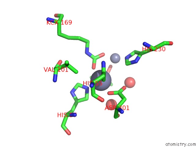
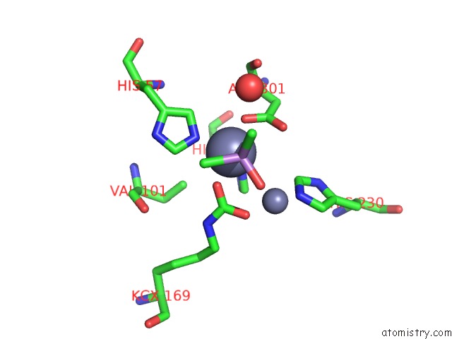
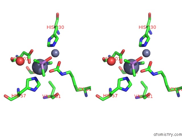
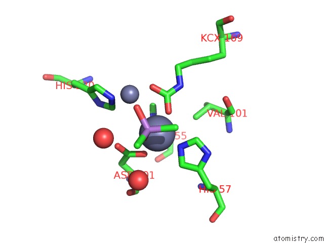
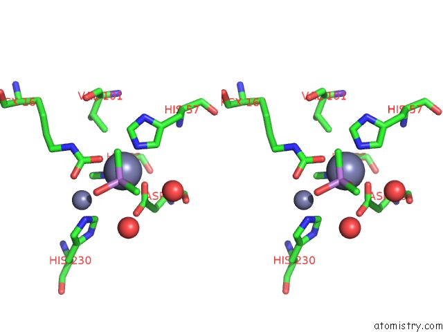
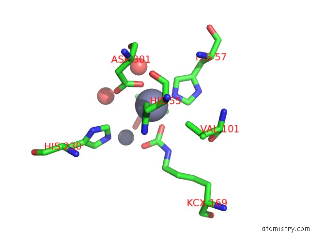
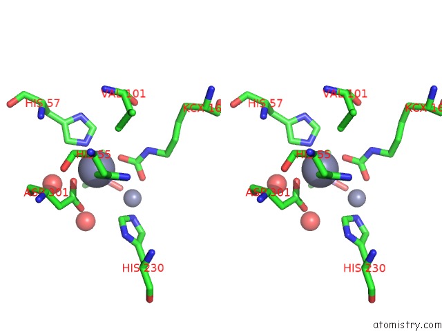
Zinc binding site 1 out of 8 in 2o4q
Go back to
Zinc binding site 1 out
of 8 in the Structure of Phosphotriesterase Mutant G60A
Mono view
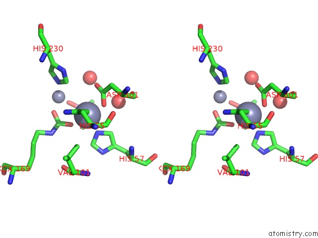
Stereo pair view
Mono view
Stereo pair view
A full contact list of Zinc with other atoms in the Zn binding
site number 1 of Structure of Phosphotriesterase Mutant G60A within 5.0Å range:
|
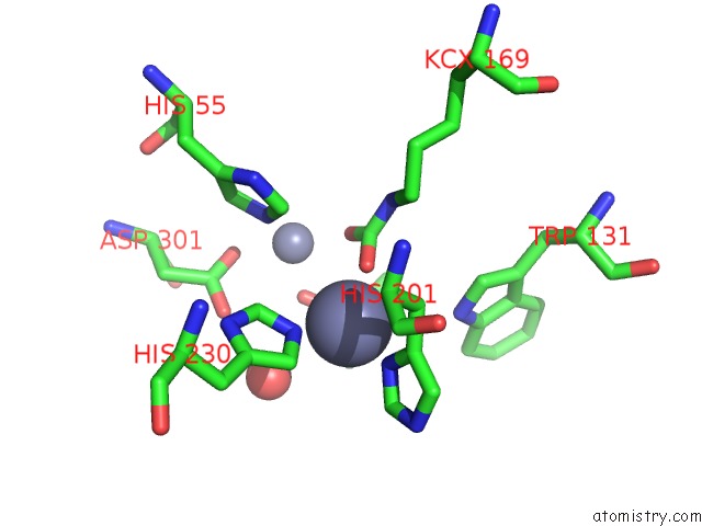
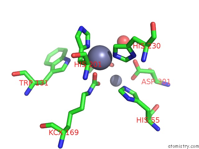
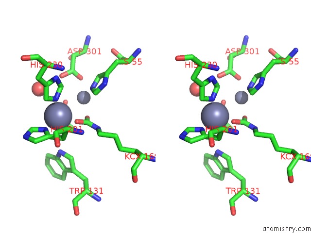
Zinc binding site 2 out of 8 in 2o4q
Go back to
Zinc binding site 2 out
of 8 in the Structure of Phosphotriesterase Mutant G60A
Mono view
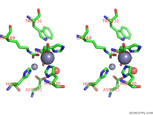
Stereo pair view
Mono view
Stereo pair view
A full contact list of Zinc with other atoms in the Zn binding
site number 2 of Structure of Phosphotriesterase Mutant G60A within 5.0Å range:
|
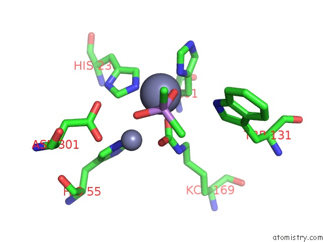
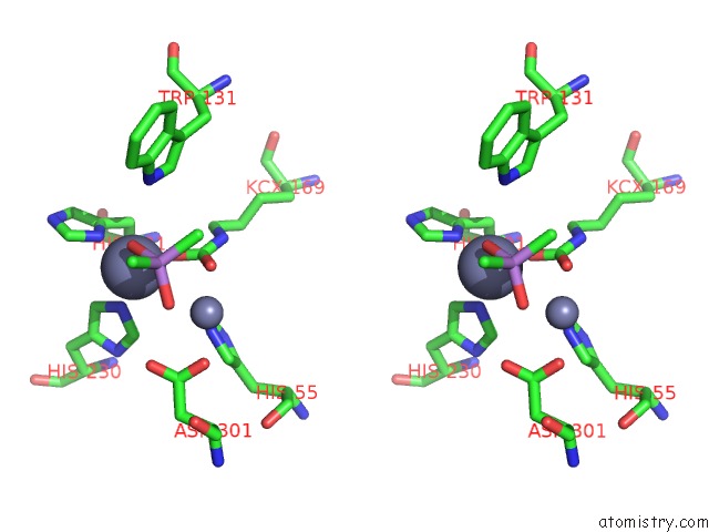
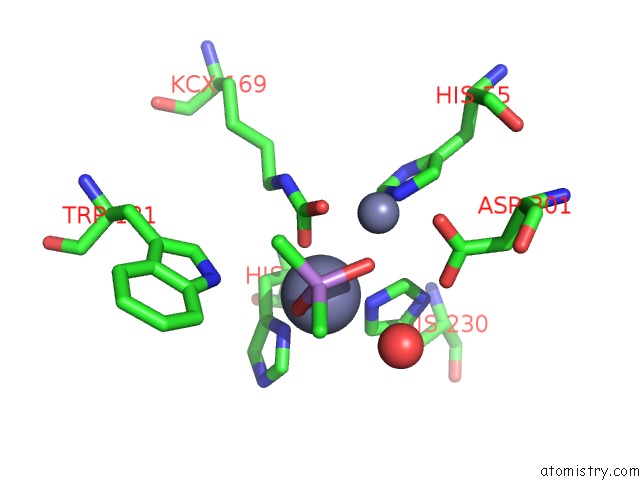
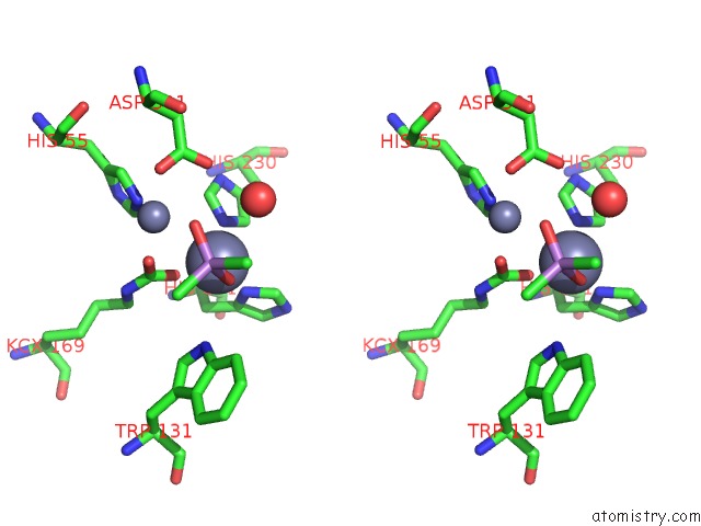
Zinc binding site 3 out of 8 in 2o4q
Go back to
Zinc binding site 3 out
of 8 in the Structure of Phosphotriesterase Mutant G60A
Mono view
Stereo pair view
Mono view
Stereo pair view
A full contact list of Zinc with other atoms in the Zn binding
site number 3 of Structure of Phosphotriesterase Mutant G60A within 5.0Å range:
|
Zinc binding site 4 out of 8 in 2o4q
Go back to
Zinc binding site 4 out
of 8 in the Structure of Phosphotriesterase Mutant G60A
Mono view
Stereo pair view
Mono view
Stereo pair view
A full contact list of Zinc with other atoms in the Zn binding
site number 4 of Structure of Phosphotriesterase Mutant G60A within 5.0Å range:
|
Zinc binding site 5 out of 8 in 2o4q
Go back to
Zinc binding site 5 out
of 8 in the Structure of Phosphotriesterase Mutant G60A
Mono view
Stereo pair view
Mono view
Stereo pair view
A full contact list of Zinc with other atoms in the Zn binding
site number 5 of Structure of Phosphotriesterase Mutant G60A within 5.0Å range:
|
Zinc binding site 6 out of 8 in 2o4q
Go back to
Zinc binding site 6 out
of 8 in the Structure of Phosphotriesterase Mutant G60A
Mono view
Stereo pair view
Mono view
Stereo pair view
A full contact list of Zinc with other atoms in the Zn binding
site number 6 of Structure of Phosphotriesterase Mutant G60A within 5.0Å range:
|
Zinc binding site 7 out of 8 in 2o4q
Go back to
Zinc binding site 7 out
of 8 in the Structure of Phosphotriesterase Mutant G60A
Mono view
Stereo pair view
Mono view
Stereo pair view
A full contact list of Zinc with other atoms in the Zn binding
site number 7 of Structure of Phosphotriesterase Mutant G60A within 5.0Å range:
|
Zinc binding site 8 out of 8 in 2o4q
Go back to
Zinc binding site 8 out
of 8 in the Structure of Phosphotriesterase Mutant G60A
Mono view
Stereo pair view
Mono view
Stereo pair view
A full contact list of Zinc with other atoms in the Zn binding
site number 8 of Structure of Phosphotriesterase Mutant G60A within 5.0Å range:
|
Reference:
J.Kim,
P.C.Tsai,
S.L.Chen,
F.Himo,
S.C.Almo,
F.M.Raushel.
Structure of Diethyl Phosphate Bound to the Binuclear Metal Center of Phosphotriesterase. Biochemistry V. 47 9497 2008.
ISSN: ISSN 0006-2960
PubMed: 18702530
DOI: 10.1021/BI800971V
Page generated: Thu Oct 17 02:26:52 2024
ISSN: ISSN 0006-2960
PubMed: 18702530
DOI: 10.1021/BI800971V
Last articles
I in 1HC0I in 1HC9
I in 1GWG
I in 1GZA
I in 1GWD
I in 1GUL
I in 1GTE
I in 1GTH
I in 1GJD
I in 1F3M