Zinc »
PDB 2mmi-2n9p »
2mvw »
Zinc in PDB 2mvw: Solution Structure of the TRIM19 B-BOX1 (B1) of Human Promyelocytic Leukemia (Pml)
Zinc Binding Sites:
The binding sites of Zinc atom in the Solution Structure of the TRIM19 B-BOX1 (B1) of Human Promyelocytic Leukemia (Pml)
(pdb code 2mvw). This binding sites where shown within
5.0 Angstroms radius around Zinc atom.
In total 4 binding sites of Zinc where determined in the Solution Structure of the TRIM19 B-BOX1 (B1) of Human Promyelocytic Leukemia (Pml), PDB code: 2mvw:
Jump to Zinc binding site number: 1; 2; 3; 4;
In total 4 binding sites of Zinc where determined in the Solution Structure of the TRIM19 B-BOX1 (B1) of Human Promyelocytic Leukemia (Pml), PDB code: 2mvw:
Jump to Zinc binding site number: 1; 2; 3; 4;
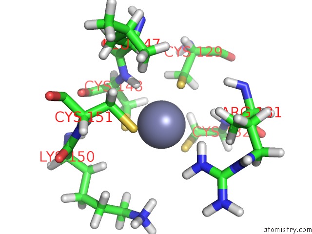
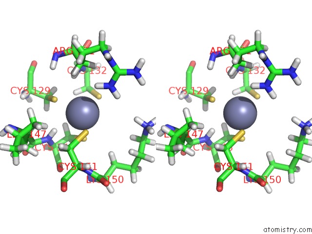
Zinc binding site 1 out of 4 in 2mvw
Go back to
Zinc binding site 1 out
of 4 in the Solution Structure of the TRIM19 B-BOX1 (B1) of Human Promyelocytic Leukemia (Pml)
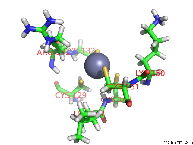
Mono view
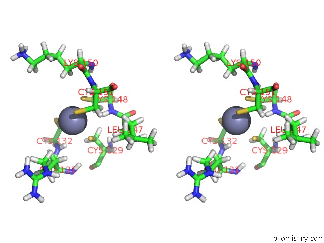
Stereo pair view
Mono view
Stereo pair view
A full contact list of Zinc with other atoms in the Zn binding
site number 1 of Solution Structure of the TRIM19 B-BOX1 (B1) of Human Promyelocytic Leukemia (Pml) within 5.0Å range:
|
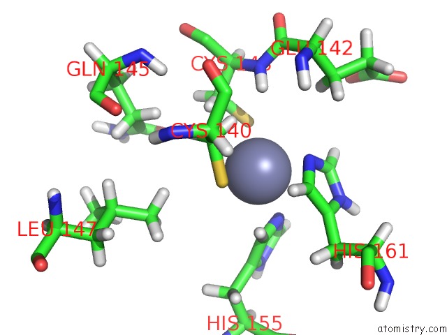
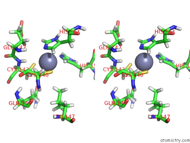
Zinc binding site 2 out of 4 in 2mvw
Go back to
Zinc binding site 2 out
of 4 in the Solution Structure of the TRIM19 B-BOX1 (B1) of Human Promyelocytic Leukemia (Pml)
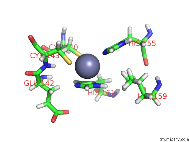
Mono view
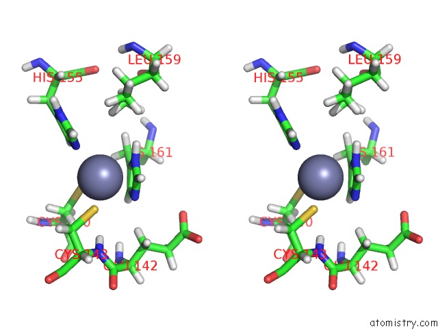
Stereo pair view
Mono view
Stereo pair view
A full contact list of Zinc with other atoms in the Zn binding
site number 2 of Solution Structure of the TRIM19 B-BOX1 (B1) of Human Promyelocytic Leukemia (Pml) within 5.0Å range:
|
Zinc binding site 3 out of 4 in 2mvw
Go back to
Zinc binding site 3 out
of 4 in the Solution Structure of the TRIM19 B-BOX1 (B1) of Human Promyelocytic Leukemia (Pml)
Mono view
Stereo pair view
Mono view
Stereo pair view
A full contact list of Zinc with other atoms in the Zn binding
site number 3 of Solution Structure of the TRIM19 B-BOX1 (B1) of Human Promyelocytic Leukemia (Pml) within 5.0Å range:
|
Zinc binding site 4 out of 4 in 2mvw
Go back to
Zinc binding site 4 out
of 4 in the Solution Structure of the TRIM19 B-BOX1 (B1) of Human Promyelocytic Leukemia (Pml)
Mono view
Stereo pair view
Mono view
Stereo pair view
A full contact list of Zinc with other atoms in the Zn binding
site number 4 of Solution Structure of the TRIM19 B-BOX1 (B1) of Human Promyelocytic Leukemia (Pml) within 5.0Å range:
|
Reference:
S.Y.Huang,
M.T.Naik,
C.F.Chang,
P.J.Fang,
Y.H.Wang,
H.M.Shih,
T.H.Huang.
The B-Box 1 Dimer of Human Promyelocytic Leukemia Protein. J.Biomol.uc(Nmr) 2014.
ISSN: ISSN 0925-2738
PubMed: 25355412
DOI: 10.1007/S10858-014-9869-4
Page generated: Thu Oct 17 02:08:38 2024
ISSN: ISSN 0925-2738
PubMed: 25355412
DOI: 10.1007/S10858-014-9869-4
Last articles
Fe in 2YXOFe in 2YRS
Fe in 2YXC
Fe in 2YNM
Fe in 2YVJ
Fe in 2YP1
Fe in 2YU2
Fe in 2YU1
Fe in 2YQB
Fe in 2YOO