Zinc »
PDB 2ics-2iv0 »
2imz »
Zinc in PDB 2imz: Crystal Structure of Mtu Reca Intein Splicing Domain
Protein crystallography data
The structure of Crystal Structure of Mtu Reca Intein Splicing Domain, PDB code: 2imz
was solved by
P.Van Roey,
with X-Ray Crystallography technique. A brief refinement statistics is given in the table below:
| Resolution Low / High (Å) | 28.76 / 1.70 |
| Space group | P 21 21 2 |
| Cell size a, b, c (Å), α, β, γ (°) | 82.430, 93.630, 40.140, 90.00, 90.00, 90.00 |
| R / Rfree (%) | 18.1 / 21 |
Zinc Binding Sites:
The binding sites of Zinc atom in the Crystal Structure of Mtu Reca Intein Splicing Domain
(pdb code 2imz). This binding sites where shown within
5.0 Angstroms radius around Zinc atom.
In total 2 binding sites of Zinc where determined in the Crystal Structure of Mtu Reca Intein Splicing Domain, PDB code: 2imz:
Jump to Zinc binding site number: 1; 2;
In total 2 binding sites of Zinc where determined in the Crystal Structure of Mtu Reca Intein Splicing Domain, PDB code: 2imz:
Jump to Zinc binding site number: 1; 2;
Zinc binding site 1 out of 2 in 2imz
Go back to
Zinc binding site 1 out
of 2 in the Crystal Structure of Mtu Reca Intein Splicing Domain
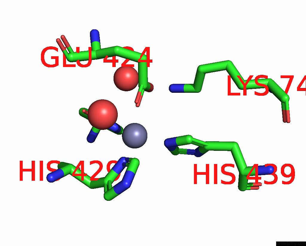
Mono view
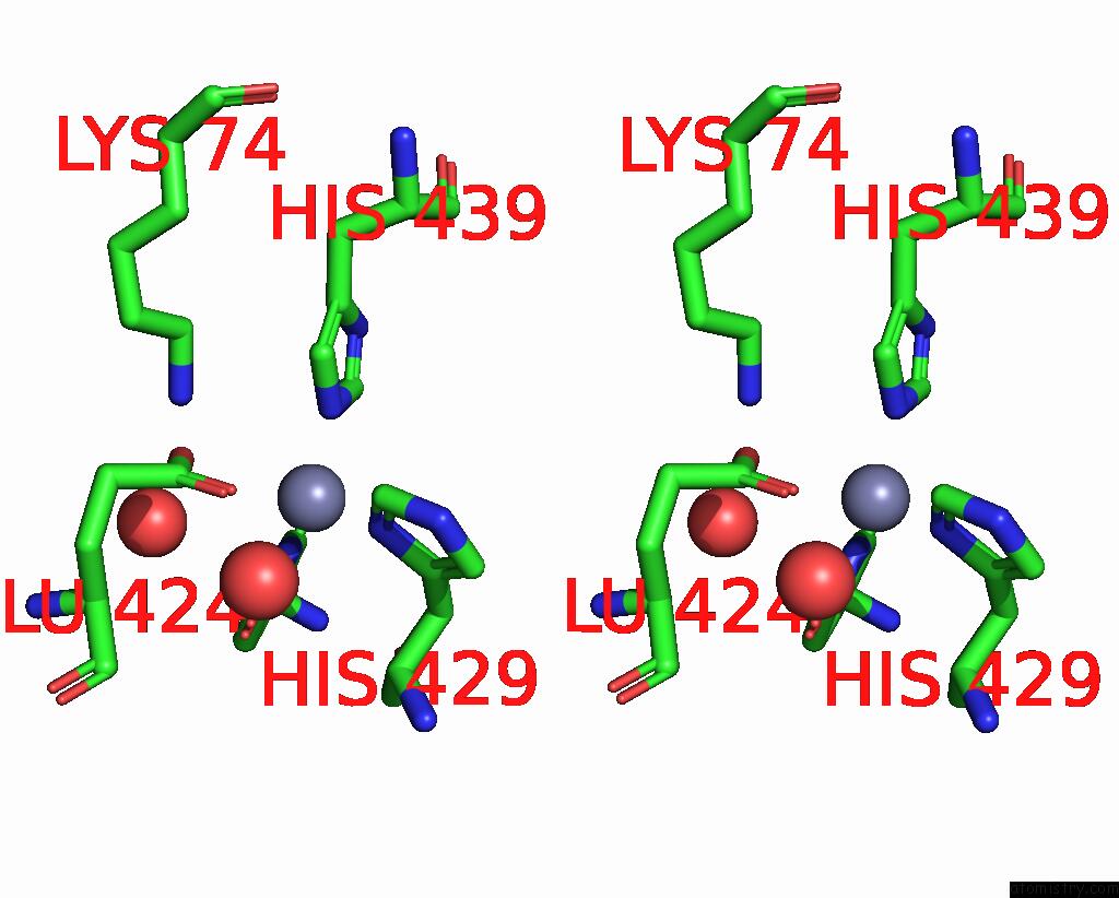
Stereo pair view
Mono view
Stereo pair view
A full contact list of Zinc with other atoms in the Zn binding
site number 1 of Crystal Structure of Mtu Reca Intein Splicing Domain within 5.0Å range:
|
Zinc binding site 2 out of 2 in 2imz
Go back to
Zinc binding site 2 out
of 2 in the Crystal Structure of Mtu Reca Intein Splicing Domain

Mono view
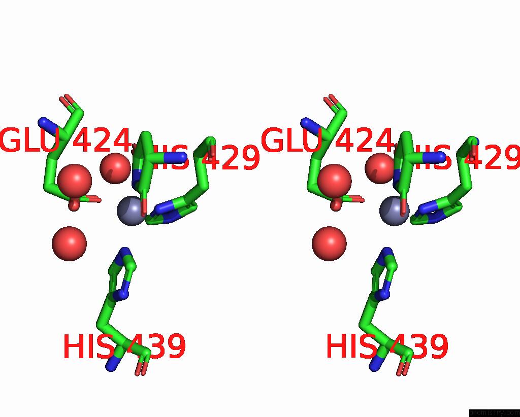
Stereo pair view
Mono view
Stereo pair view
A full contact list of Zinc with other atoms in the Zn binding
site number 2 of Crystal Structure of Mtu Reca Intein Splicing Domain within 5.0Å range:
|
Reference:
P.Van Roey,
B.Pereira,
Z.Li,
K.Hiraga,
M.Belfort,
V.Derbyshire.
Crystallographic and Mutational Studies of Mycobacterium Tuberculosis Reca Mini-Inteins Suggest A Pivotal Role For A Highly Conserved Aspartate Residue. J.Mol.Biol. V. 367 162 2007.
ISSN: ISSN 0022-2836
PubMed: 17254599
DOI: 10.1016/J.JMB.2006.12.050
Page generated: Thu Oct 17 00:57:00 2024
ISSN: ISSN 0022-2836
PubMed: 17254599
DOI: 10.1016/J.JMB.2006.12.050
Last articles
Mg in 6QCSMg in 6QBV
Mg in 6QBD
Mg in 6QBT
Mg in 6Q84
Mg in 6Q8C
Mg in 6Q8B
Mg in 6Q82
Mg in 6Q89
Mg in 6Q8A