Zinc »
PDB 2ics-2iv0 »
2imt »
Zinc in PDB 2imt: The X-Ray Structure of A Bak Homodimer Reveals An Inhibitory Zinc Binding Site
Protein crystallography data
The structure of The X-Ray Structure of A Bak Homodimer Reveals An Inhibitory Zinc Binding Site, PDB code: 2imt
was solved by
T.Moldoveanu,
Q.Liu,
A.Tocilj,
M.Watson,
G.C.Shore,
K.B.Gehring,
with X-Ray Crystallography technique. A brief refinement statistics is given in the table below:
| Resolution Low / High (Å) | 37.32 / 1.49 |
| Space group | C 1 2 1 |
| Cell size a, b, c (Å), α, β, γ (°) | 57.247, 53.646, 58.074, 90.00, 114.87, 90.00 |
| R / Rfree (%) | 19.3 / 22 |
Zinc Binding Sites:
The binding sites of Zinc atom in the The X-Ray Structure of A Bak Homodimer Reveals An Inhibitory Zinc Binding Site
(pdb code 2imt). This binding sites where shown within
5.0 Angstroms radius around Zinc atom.
In total only one binding site of Zinc was determined in the The X-Ray Structure of A Bak Homodimer Reveals An Inhibitory Zinc Binding Site, PDB code: 2imt:
In total only one binding site of Zinc was determined in the The X-Ray Structure of A Bak Homodimer Reveals An Inhibitory Zinc Binding Site, PDB code: 2imt:
Zinc binding site 1 out of 1 in 2imt
Go back to
Zinc binding site 1 out
of 1 in the The X-Ray Structure of A Bak Homodimer Reveals An Inhibitory Zinc Binding Site
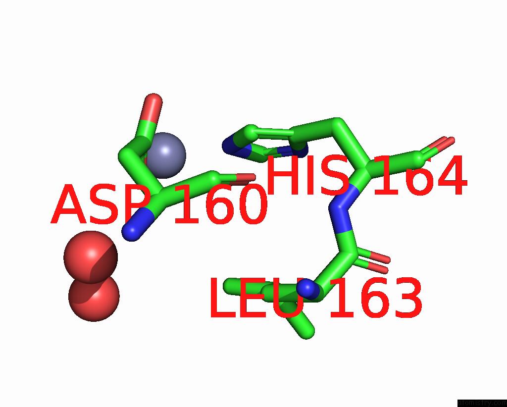
Mono view
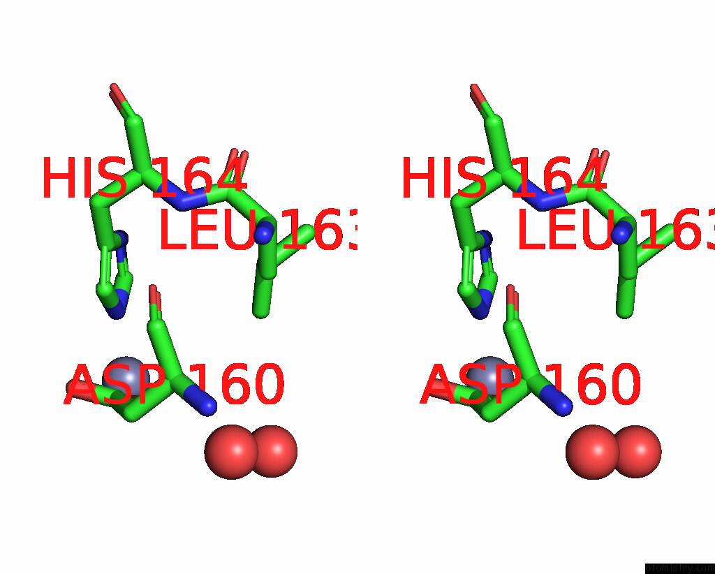
Stereo pair view
Mono view
Stereo pair view
A full contact list of Zinc with other atoms in the Zn binding
site number 1 of The X-Ray Structure of A Bak Homodimer Reveals An Inhibitory Zinc Binding Site within 5.0Å range:
|
Reference:
T.Moldoveanu,
Q.Liu,
A.Tocilj,
M.Watson,
G.Shore,
K.Gehring.
The X-Ray Structure of A Bak Homodimer Reveals An Inhibitory Zinc Binding Site. Mol.Cell V. 24 677 2006.
ISSN: ISSN 1097-2765
PubMed: 17157251
DOI: 10.1016/J.MOLCEL.2006.10.014
Page generated: Thu Oct 17 00:56:39 2024
ISSN: ISSN 1097-2765
PubMed: 17157251
DOI: 10.1016/J.MOLCEL.2006.10.014
Last articles
Mg in 6SYUMg in 6SYT
Mg in 6SYR
Mg in 6SY1
Mg in 6SWL
Mg in 6SWE
Mg in 6SUL
Mg in 6SUR
Mg in 6SVP
Mg in 6SVM