Zinc »
PDB 2hqk-2ibi »
2hvf »
Zinc in PDB 2hvf: Crystal Structure of N-Terminal Domain of Ribosomal Protein L9 (NTL9), G34DA
Protein crystallography data
The structure of Crystal Structure of N-Terminal Domain of Ribosomal Protein L9 (NTL9), G34DA, PDB code: 2hvf
was solved by
B.Anil,
E.Y.Kim,
J.H.Cho,
H.Schindelin,
D.P.Raleigh,
with X-Ray Crystallography technique. A brief refinement statistics is given in the table below:
| Resolution Low / High (Å) | 20.00 / 1.57 |
| Space group | P 41 21 2 |
| Cell size a, b, c (Å), α, β, γ (°) | 53.517, 53.517, 36.178, 90.00, 90.00, 90.00 |
| R / Rfree (%) | 15.9 / 20.8 |
Other elements in 2hvf:
The structure of Crystal Structure of N-Terminal Domain of Ribosomal Protein L9 (NTL9), G34DA also contains other interesting chemical elements:
| Chlorine | (Cl) | 5 atoms |
Zinc Binding Sites:
The binding sites of Zinc atom in the Crystal Structure of N-Terminal Domain of Ribosomal Protein L9 (NTL9), G34DA
(pdb code 2hvf). This binding sites where shown within
5.0 Angstroms radius around Zinc atom.
In total 4 binding sites of Zinc where determined in the Crystal Structure of N-Terminal Domain of Ribosomal Protein L9 (NTL9), G34DA, PDB code: 2hvf:
Jump to Zinc binding site number: 1; 2; 3; 4;
In total 4 binding sites of Zinc where determined in the Crystal Structure of N-Terminal Domain of Ribosomal Protein L9 (NTL9), G34DA, PDB code: 2hvf:
Jump to Zinc binding site number: 1; 2; 3; 4;
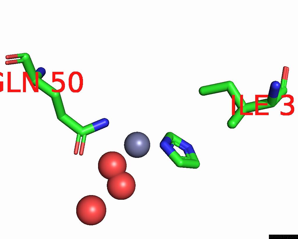
Zinc binding site 1 out of 4 in 2hvf
Go back to
Zinc binding site 1 out
of 4 in the Crystal Structure of N-Terminal Domain of Ribosomal Protein L9 (NTL9), G34DA
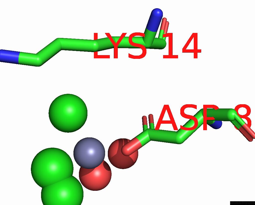
Mono view
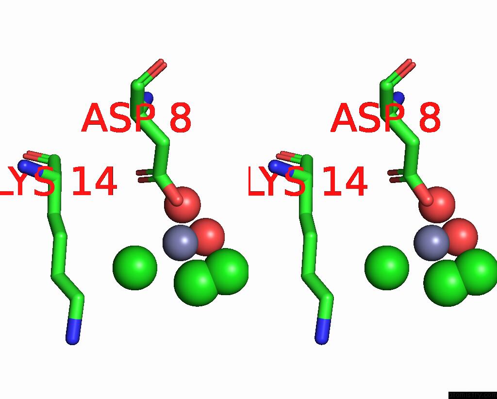
Stereo pair view
Mono view
Stereo pair view
A full contact list of Zinc with other atoms in the Zn binding
site number 1 of Crystal Structure of N-Terminal Domain of Ribosomal Protein L9 (NTL9), G34DA within 5.0Å range:
|
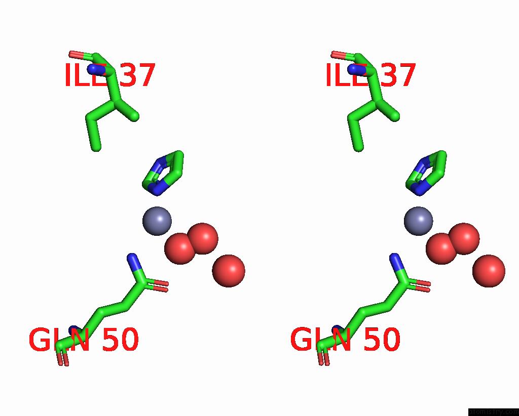
Zinc binding site 2 out of 4 in 2hvf
Go back to
Zinc binding site 2 out
of 4 in the Crystal Structure of N-Terminal Domain of Ribosomal Protein L9 (NTL9), G34DA
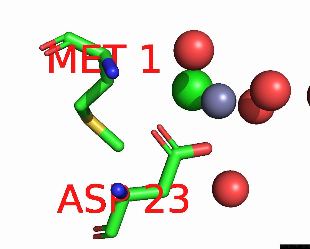
Mono view
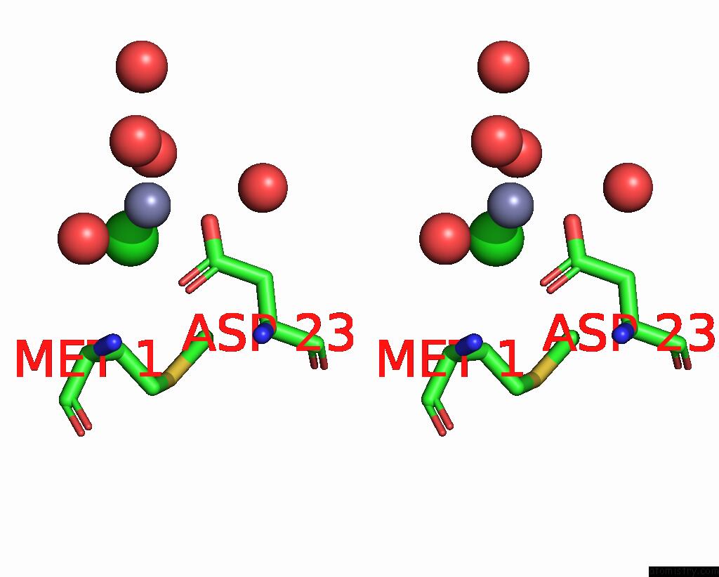
Stereo pair view
Mono view
Stereo pair view
A full contact list of Zinc with other atoms in the Zn binding
site number 2 of Crystal Structure of N-Terminal Domain of Ribosomal Protein L9 (NTL9), G34DA within 5.0Å range:
|
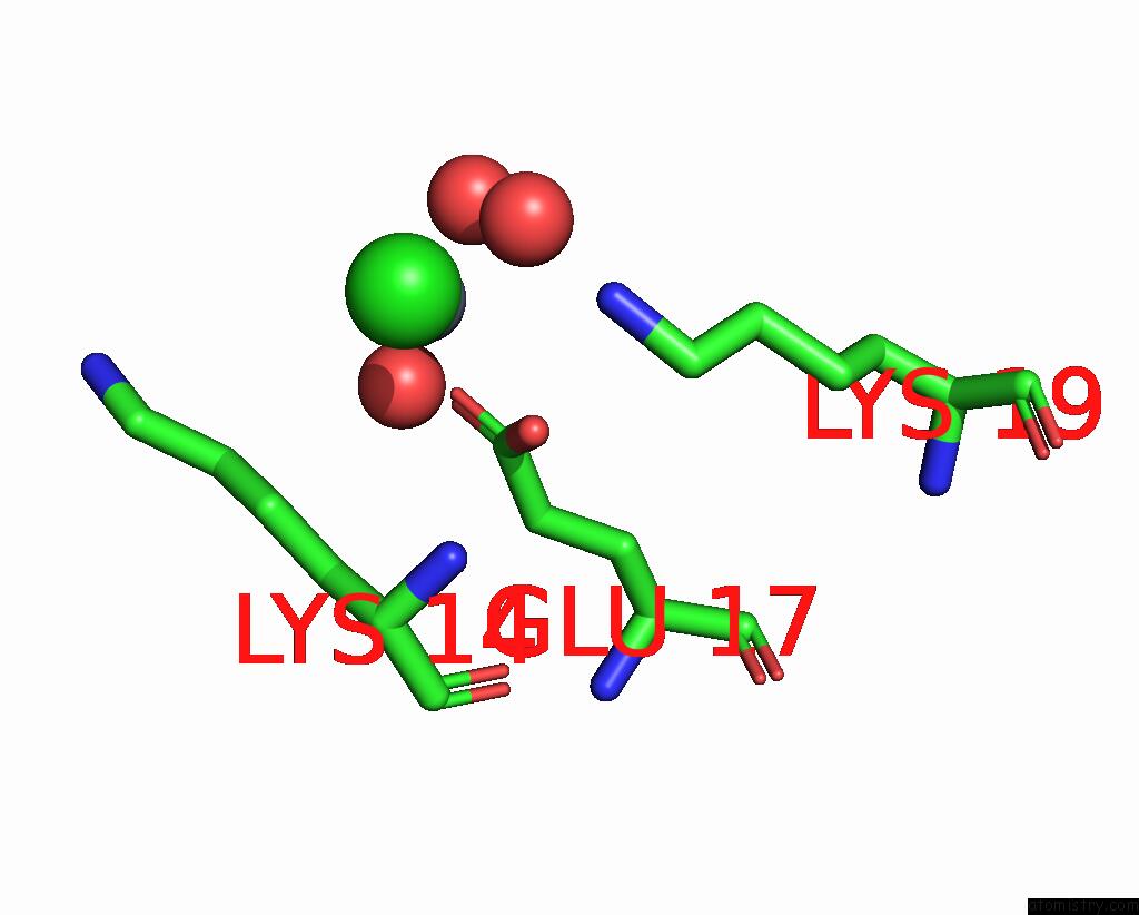
Zinc binding site 3 out of 4 in 2hvf
Go back to
Zinc binding site 3 out
of 4 in the Crystal Structure of N-Terminal Domain of Ribosomal Protein L9 (NTL9), G34DA
Mono view
Stereo pair view
Mono view
Stereo pair view
A full contact list of Zinc with other atoms in the Zn binding
site number 3 of Crystal Structure of N-Terminal Domain of Ribosomal Protein L9 (NTL9), G34DA within 5.0Å range:
|
Zinc binding site 4 out of 4 in 2hvf
Go back to
Zinc binding site 4 out
of 4 in the Crystal Structure of N-Terminal Domain of Ribosomal Protein L9 (NTL9), G34DA
Mono view

Stereo pair view
Mono view
Stereo pair view
A full contact list of Zinc with other atoms in the Zn binding
site number 4 of Crystal Structure of N-Terminal Domain of Ribosomal Protein L9 (NTL9), G34DA within 5.0Å range:
|
Reference:
B.Anil,
E.Y.Kim,
J.H.Cho,
H.Schindelin,
D.P.Raleigh.
Detecting and Quantifying Strain in Protein Folding To Be Published.
Page generated: Wed Aug 20 03:25:32 2025
Last articles
Mn in 9LJUMn in 9LJW
Mn in 9LJS
Mn in 9LJR
Mn in 9LJT
Mn in 9LJV
Mg in 9UA2
Mg in 9R96
Mg in 9VM1
Mg in 9P01