Zinc »
PDB 2geh-2gzg »
2giv »
Zinc in PDB 2giv: Human Myst Histone Acetyltransferase 1
Enzymatic activity of Human Myst Histone Acetyltransferase 1
All present enzymatic activity of Human Myst Histone Acetyltransferase 1:
2.3.1.48;
2.3.1.48;
Protein crystallography data
The structure of Human Myst Histone Acetyltransferase 1, PDB code: 2giv
was solved by
J.Min,
H.Wu,
P.Loppnau,
J.Weigelt,
M.Sundstrom,
C.H.Arrowsmith,
A.M.Edwards,
A.Bochkarev,
A.N.Plotnikov,
Structural Genomicsconsortium (Sgc),
with X-Ray Crystallography technique. A brief refinement statistics is given in the table below:
| Resolution Low / High (Å) | 41.89 / 1.94 |
| Space group | P 21 21 21 |
| Cell size a, b, c (Å), α, β, γ (°) | 46.305, 58.577, 119.753, 90.00, 90.00, 90.00 |
| R / Rfree (%) | 20.7 / 26.2 |
Other elements in 2giv:
The structure of Human Myst Histone Acetyltransferase 1 also contains other interesting chemical elements:
| Chlorine | (Cl) | 1 atom |
Zinc Binding Sites:
The binding sites of Zinc atom in the Human Myst Histone Acetyltransferase 1
(pdb code 2giv). This binding sites where shown within
5.0 Angstroms radius around Zinc atom.
In total only one binding site of Zinc was determined in the Human Myst Histone Acetyltransferase 1, PDB code: 2giv:
In total only one binding site of Zinc was determined in the Human Myst Histone Acetyltransferase 1, PDB code: 2giv:
Zinc binding site 1 out of 1 in 2giv
Go back to
Zinc binding site 1 out
of 1 in the Human Myst Histone Acetyltransferase 1

Mono view
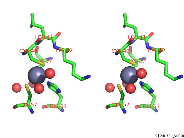
Stereo pair view
Mono view
Stereo pair view
A full contact list of Zinc with other atoms in the Zn binding
site number 1 of Human Myst Histone Acetyltransferase 1 within 5.0Å range:
|
Reference:
H.Wu,
J.Min,
P.Loppnau,
J.Weigelt,
M.Sundstrom,
C.H.Arrowsmith,
A.M.Edwards,
A.Bochkarev,
A.N.Plotnikov.
The Crystal Structure of Human Myst Histone Acetyltransferase 1 in Complex with Acetylcoenzyme A To Be Published.
Page generated: Thu Oct 17 00:19:19 2024
Last articles
Mg in 5NH6Mg in 5NHZ
Mg in 5NH5
Mg in 5NH4
Mg in 5NG1
Mg in 5N9Z
Mg in 5NH2
Mg in 5NFZ
Mg in 5NFV
Mg in 5NG0