Zinc »
PDB 2fos-2g2p »
2g02 »
Zinc in PDB 2g02: Nisin Cyclase
Protein crystallography data
The structure of Nisin Cyclase, PDB code: 2g02
was solved by
S.K.Nair,
with X-Ray Crystallography technique. A brief refinement statistics is given in the table below:
| Resolution Low / High (Å) | 20.00 / 2.50 |
| Space group | I 41 |
| Cell size a, b, c (Å), α, β, γ (°) | 154.941, 154.941, 50.956, 90.00, 90.00, 90.00 |
| R / Rfree (%) | 18.1 / 24.2 |
Zinc Binding Sites:
The binding sites of Zinc atom in the Nisin Cyclase
(pdb code 2g02). This binding sites where shown within
5.0 Angstroms radius around Zinc atom.
In total only one binding site of Zinc was determined in the Nisin Cyclase, PDB code: 2g02:
In total only one binding site of Zinc was determined in the Nisin Cyclase, PDB code: 2g02:
Zinc binding site 1 out of 1 in 2g02
Go back to
Zinc binding site 1 out
of 1 in the Nisin Cyclase
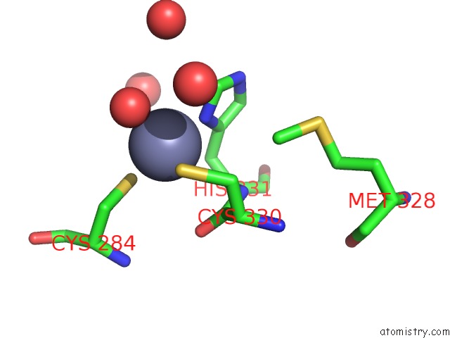
Mono view
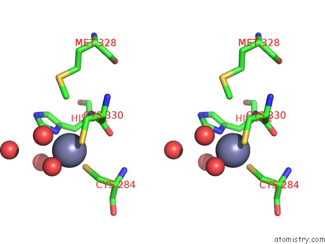
Stereo pair view
Mono view
Stereo pair view
A full contact list of Zinc with other atoms in the Zn binding
site number 1 of Nisin Cyclase within 5.0Å range:
|
Reference:
B.Li,
J.P.J.Yu,
J.S.Brunzelle,
G.N.Moll,
W.A.Van Der Donk,
S.K.Nair.
Structure and Mechanism of the Lantibiotic Cyclase Involved in Nisin Biosynthesis Science V. 311 1464 2006.
ISSN: ISSN 0036-8075
PubMed: 16527981
DOI: 10.1126/SCIENCE.1121422
Page generated: Wed Oct 16 23:59:41 2024
ISSN: ISSN 0036-8075
PubMed: 16527981
DOI: 10.1126/SCIENCE.1121422
Last articles
Mn in 2GTXMn in 2GNM
Mn in 2GND
Mn in 2GMV
Mn in 2GLF
Mn in 2GLK
Mn in 2GDS
Mn in 2G50
Mn in 2GDF
Mn in 2G8I